
Chapter 1
Introduction
The unique characteristics of nanofibers, which have a high porosity, and superior mechanical, high surface area, electrical, and chemical capabilities, have caused them to become significant in a variety of applications at this time [1]. With time, attention on nanofibers has grown, and a process for producing them has been created [2]. Numerous methods have been used to create nanofibers; these methods can be divided into two categories based on how the nanofibers were created, namely electrospinning methods and non-electrospinning methods. When deciding between these approaches, it is important to take into account the desired alignment, the price, the quantity, and the amount of fibrous material required. These methods could be used to create various nanofiber architectures, such as core-shell, bicomponent, hollow, and porous structures [3]. Coaxial electrospinning, for instance, can be used to create core-sheath structures. Direct coaxial spinning, electrospinning, or chemical vapour deposition (CVD) can be used to create hollow nanofibers. Phase separation can be used to create porous nanofibers. The most popular technique for creating nanofibers is electrospinning, often known as electrostatic spinning [4]. Using this mechanical and electrical technology and a high-voltage power source, it is possible to create fibres with a submicron diameter [5]. The most popular method for creating nanofibers, electrospinning, has been studied by numerous researchers, and its fundamental design has been changed over time to improve productivity and get around some of its drawbacks. As a result, there are numerous subtypes of electrospinning technique. Electrospun polymeric nanofibers have distinguished themselves as a fascinating and adaptable class of materials with significant potential for biological applications among the wide variety of nanomaterials and nanostructures [6]. Researchers and professionals in the biomedical sector have paid close attention to these nanofibers because of their high surface area-to-volume ratio, adjustable characteristics, and biocompatibility [7]. The early 20th century electrospinning technology has developed into a very adaptable and effective method for creating ultrafine fibres at the nanoscale [8]. With this technique, continuous nanofibers with precisely designed diameter, composition, and morphology are created by carefully controlling the deposition of polymer solutions or melts through an electrically charged jet [9]. The distinctive electrospinning method makes it possible to create nanofibers from a wide range of biocompatible and biodegradable polymers, providing a wide range of options for adjusting their properties to particular biological needs. Biomedical research and electrospun polymeric nanofibers are a potential and fascinating nexus [10]. These nanofibers have outstanding qualities that make them the perfect choice for a wide range of biomedical applications, including tissue engineering, drug transport, wound healing, and diagnostic platforms. Additionally, their fibrous shape closely resembles the extracellular matrix present in real tissues, enabling cell adhesion, proliferation, and differentiation. Their high surface area facilitates effective drug loading and regulated release. Additionally, the electrospinning procedure enables the inclusion of bioactive substances into the nanofibers, improving their therapeutic potential. These substances include growth factors and antibacterial substances [11]. Electrospun polymeric nanofibers have distinguished themselves as a particularly promising and adaptable class of materials with exceptional potential in the area of biomedical applications among the countless nanomaterials at the forefront of this revolution.
The goal of this study is to present a thorough overview of the creation and biomedical uses of electrospun polymeric nanofibers. We will examine the different factors that affect the form and characteristics of nanofibers as we delve into the core concepts of electrospinning. We will also emphasise how crucial material choice and functionalization are in modifying electrospun nanofibers for certain biomedical applications. Additionally, we will look at recent developments in the application of electrospun nanofibers in tissue engineering, drug delivery, wound healing, and diagnostics, highlighting their potential to treat some of the most urgent problems in contemporary medicine. We invite readers to discover the fascinating advancements and game-changing potential of these nanomaterials in enhancing human health and wellbeing as we set off on this voyage into the realm of electrospun polymeric nanofibers and their medicinal applications.
1.1 Electrospinning
The technique that is most frequently utilised to create nanofibers is electrospinning. A high-voltage source, a capillary tube with a small-diameter pipette or needle, and a metal-collecting screen are tools required for electrospinning. The polymer solution is where one electrode is inserted, and the collector is where the other electrode is attached [12]. The end of the capillary tube holding the polymer solution is subjected to an electric field, which creates a charge on the liquid’s surface [13]. The fluid’s hemispherical surface at the end of the capillary tube elongates into a conical shape known as the Taylor cone as the strength of the electric field increases. When the electric field is increased further, a critical value is reached where the charged jet of fluid is ejected from the Taylor cone’s tip because the repelling electrostatic force has defeated the surface tension. Because the discharged polymer solution jet is unstable, it elongates and grows incredibly long and thin. With the evaporation of the solvent, charged polymer fibres solidify. Nanofibers are gathered in an erratic pattern on the collector [14]. The use of specialised collectors, such as the spinning drum, metal frame, or two-parallel plates system, can also collect nanofibers in a highly aligned manner [15]. To create nanofibers with homogeneous diameters and morphologies, it is necessary to regulate variables like polymer content and jet stream velocity. Numerous polymer varieties are converted into nanofibers using the electrospinning technology. The extracellular matrix (ECM) and a network of electrospun nanofibers are closely related. This similarity is a key benefit of electrospinning since it allows for the imitation of the ECM’s mechanical characteristics, high porosity, and fibre sizes. Further advancements in electrospinning are being made in order to produce continuous nanofibers in large quantities.
An electrostatically driven jet of polymer solution is used in the electrospinning process to create polymer nanofibers [16]. Over the past few years, this field has seen significant advancements, and a variety of uses for this technology have been discovered. The majority of recent research on electrospinning has either concentrated on finding the ideal conditions for electrospinning different polymers and biopolymers or on trying to understand deeper changes to many fundamental aspects of the process in order to gain control of nanofibre morphology, structure, surface functionality, and assembly strategies. Even after being electrospun, the nanofibres can be positioned in order to create specialised functional nanostructures like nanotubes and nanowires. A wide variety of fabric qualities, including strength, weight and porosity, surface functioning, etc., can also be accomplished based on the particular polymer being utilised. A variety of polymers, fibres, and particles can be laced together using this innovative fibre spinning technology to create ultra-thin layers. The dry nanofibers can contain small, insoluble particles that have been dissolved in the polymer solution [17]. Non-woven mats can have soluble medications or microbes electrospun into them. Given that they have diameters in the nanometre range and lengths of many metres, nanofibres serve as a bridge between the nanoscale and macroscale worlds. Therefore, the focus of current research is to take advantage of these properties and concentrate on figuring out the best conditions for electrospinning different polymers and biopolymers for potential applications, such as multifunctional membranes, structural components for biomedical devices, protective shields in specialty fabrics, filter media for submicron particles in the separation industry, composite reinforcement, and structures for nano-electronic machines. The majority of recent electrospinning research has focused on the many polymer-solvent solutions that can be used to create fibres. A few studies have also directly or indirectly addressed the processing/property correlations in electrospun polymer fibres. The flow rate of the solution, polymer content, molecular weight, and the distance from the syringe needle tip to the ground collecting plate have all been taken into account as processing parameters for nanofibres. It has been discovered that solution viscosity affects the fibre diameter, droplet shape at the beginning, and jet trajectory. Larger diameter fibre synthesis has been linked to increasing solution viscosity [18].
1.2 Fundamentals of Electrospinning
Electrospinning is the process used most frequently to create nanofibers. A flexible and popular method for creating nanofibers from polymer solutions or melts is electrospinning [19]. Depending on how the polymer is made, the electrospinning process can be divided into two groups: melt electrospinning and solution electrospinning. In solution electrospinning, several researchers have carefully examined the aspects influencing the electrospun web’s characteristics, distinctive traits, and diverse uses. Despite numerous attempts, the solution electrospinning process has many flaws, including low productivity, the need for an additional solvent extraction procedure, and environmental problems due to the use of harmful solvents. Although the same limitations do not apply to the melt electrospinning technique, little research has been done on it. This is due to the challenges involved in the production of finer fibres, the increased viscosity of molten polymer, and the issue of electrical discharge brought on by applying high voltage to a polymer melt [20].
The creation of a polymer solution kicks off the procedure. A polymer is dissolved in an appropriate solvent in this stage to produce a homogenous solution. The preferred characteristics of the nanofibers, such as biocompatibility, mechanical strength, and degradation rate, influence the choice of polymer and solvent. A small-diameter metal or polymer needle, known as a spinneret, is affixed to the bottom of the reservoir after the polymer solution has been poured into a syringe or reservoir. The electrospinning procedure occurs at the spinneret’s tip. The spinneret has a high-voltage power supply attached. The droplet of polymer solution is surrounded by an electric field when the voltage is introduced. The droplet’s surface develops charges as a result of this electric field. The repelling forces between like charges on the surface of the droplet become stronger than the surface tension of the polymer solution as the electric field strength increases. At the apex of the spinneret, this causes the Taylor cone, a sharp, conical structure, to form. A fine, continuous jet of the polymer solution is ejected from the tip of the Taylor cone after the electric field exceeds the surface tension. The jet is directed towards a collector that is grounded or oppositely charged and is placed a specified distance away. Due to its typically low boiling point, the solvent in the ejected jet rapidly evaporates as it travels in the direction of the collector. Solid nanofibers are produced as a result of the polymer solidifying as the solvent evaporates. On the grounded or negatively charged collector, the solid nanofibers are gathered. Depending on the intended nanofiber alignment and layout, the collector may take the shape of a revolving drum, a stationary plate, or another arrangement. The morphology, diameter, mechanical strength, and chemical content of the electrospun nanofibers can all be determined once they have been collected. Characterization often involves the use of methods like spectroscopy, atomic force microscopy, and scanning electron microscopy (SEM).
The exquisite control over nanofiber diameter, porosity, and alignment provided by the electrospinning technique makes it appropriate for a variety of applications. Researchers can modify the nanofiber qualities for particular uses, such as drug delivery systems, tissue engineering scaffolds, wound dressings, and more by adjusting process parameters including voltage, flow rate, and collector distance.
1.3 Fabrication of Electrospun Polymer Nanofibers
The manufacture of ultrafine fibres with dimensions ranging from the nanometer to the micrometre range is made possible by the fabrication of electrospun polymer nanofibers, a highly adaptable and popular manufacturing technology. This technology has shown to be important in a variety of sectors, including filtration, biomedicine, materials science, and environmental science [21]. Considering the desired qualities of the nanofibers, a suitable polymer is carefully chosen to start the process. Polymers come in a variety of types that are often used, from traditional materials like polyethylene and polyurethane to biodegradable possibilities like poly (lactic acid) (PLA) and poly (glycolic acid) (PGA). In order to create a homogeneous solution, the selected polymer is then dissolved in a compatible solvent. The next stage includes setting up a spinneret to extrude the polymer solution. A spinneret typically consists of a thin needle or nozzle. A conical structure known as the Taylor cone is formed when a spinneret is subjected to a high-voltage, which causes an electric field to induce charges on the droplet of polymer solution. A fine jet of the polymer solution is released from the tip of the Taylor cone and is directed in the direction of a collector when the electric field reaches a critical strength [22]. The jet’s solvent quickly evaporates during this flight, leaving behind solidified nanofibers. The alignment and arrangement of the nanofibers can be controlled by placing the collected nanofibers on a grounded or oppositely charged surface. Using methods like scanning electron microscopy (SEM) and spectroscopy, further characterization of the electrospun nanofibers includes evaluations of morphology, diameter, mechanical properties, and chemical composition. Overall, the production of electrospun polymer nanofibers offers a crucial tool for modifying the characteristics of nanofibers to meet particular commercial and scientific difficulties across a range of applications. These days, in addition to polymers, electrospun micro/nanofibers are also based on ceramics, metals, metal oxides, organic composite systems, and inorganic composite systems. These copolymer, mix, or organic filler-based electrospun membranes consistently display improved mechanical behaviour, barrier characteristics, and thermal stability. Drug-containing electrospun fibre mats have drawn a lot of attention for use in a variety of biomedical applications, such as tissue regeneration, wound dressing, and the prevention of anaerobic bacterial colonisation, among others. Fibrous carriers are more promising than microspheres, hydrogels, and micelle systems because of their versatile adaptability and comparative simplicity of application.
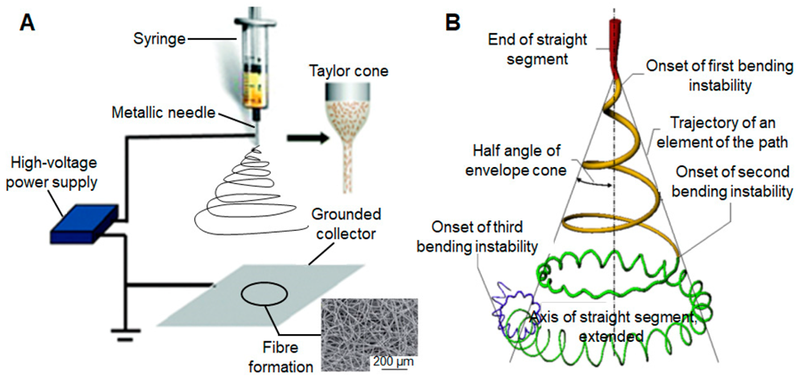
A high-voltage source that creates an electrical field between a positively-charged syringe needle and a grounded collector, a metallic needle where the charged solution is forced to stretch due to electrostatic forces, a syringe pump, and a grounded target to deposit the resulting fibres make up the main electrospinning apparatus. The metallic needle is connected to the high-power supply via electrical cables, which also maintains a close proximity between the target and the syringe tube. Electrostatic repulsion will cause the jet to lengthen as the solvent evaporates while the electrode is electrospun. A uniform fibre is formed in the micro- to nanoscale as a result of the process of thinning, which comes next. This fibre can be gathered in a variety of orientations to produce specialised structures with various chemical compositions and mechanical qualities. Numerous targets, mostly copper plates, rotating drums, and aluminium foil, have been used to gather fibres during electrospinning up to this point. More than 40 distinct types of natural and synthetic organic polymers have already been successfully electrospun into fibres with diameters ranging from tens of nanometers to a few micrometres with the use of this comparatively simple process.
1.4 Polymer Nanofibres for Biomedical Applications
Fibres with a diameter in the nanometre range are known as nanofibres. Nanofibers can be produced from a variety of polymers, giving them a range of physical characteristics and possible uses [23]. Collagen, cellulose, silk fibroin, keratin, gelatin, and polysaccharides like chitosan and alginate are a few examples of natural polymers. Poly(3-hydroxybutyrate-co-3-hydroxyvalerate), Poly (lactic acid), polycaprolactone (PCL), poly (lactic-co-glycolic acid), polyurethane (PU), and poly (ethylene-co-vinyl acetate) are a few examples of synthetic polymers. Covalent bonds are used to join polymer chains together. The type of polymer utilised and the manufacturing process affect the nanofibers’ sizes. When compared to their microfiber counterparts, polymer nanofibers have a greater surface area-to-volume ratio, higher porosity, considerable mechanical strength, and greater flexibility in functionalization. Nanofibers can be produced using a wide range of techniques, including drawing, electrospinning, self-assembly, template synthesis, and thermally induced phase separation. The most popular technique for creating nanofibers is electrospinning, which has a simple setup, the capacity to mass-produce continuous nanofibers from a variety of polymers, and the ability to create ultrathin fibres with adjustable diameters, compositions, and orientations. This flexibility enables the fibres’ shape and arrangement to be controlled, enabling the fabrication of various structures according to the application goals.
The majority of human organs and tissues, including bone, collagen, dentin, and skin, are nanofibrous in nature. Their organised hierarchical fibre structures, which realign at the nanoscale, are what distinguish them and drive the majority of nanofiber research towards biological and bioengineering applications. Fibres and fibre mats have peculiar physical characteristics, such as surface area, diameter, and porosity, that are similar to those of the extracellular matrix (ECM). By separating tissues from one another and providing potential anchoring and support for cells, this new shape improves cell behaviour. The functional features of biomaterials may be exceptionally well suited to ECM characteristics. Fibre mats are employed to regulate several characteristics of cell tissue, including cell migration and proliferation. Other biomedical applications for nanofibrous materials include dental materials, scaffolds for enzyme immobilisation, medical implants, wound dressings, antibacterial agents, drug delivery systems, biomimetic actuators, and protective textiles against biological and chemical hazards. Nanofibers utilised in biomedical applications must be made from biocompatible and biodegradable polymers.
1.4.1 Tissue Engineering
Tissue engineering is not just restricted to applications involving cells and tissue scaffolds; it also includes the utilisation of cells positioned on tissue scaffolds in the production of new living tissue for medicinal purposes [24]. It was originally thought of as a sub-field of biomaterials, but since it has expanded in importance and scope, it can now be viewed as a separate field. The phrase has also been used to describe attempts to carry out particular biochemical processes using cells from a support system that has been artificially generated. Although the terms “regenerative medicine” and “tissue engineering” are frequently used interchangeably, individuals who practise regenerative medicine emphasise the utilisation of stem cells or progenitor cells more than tissue engineering.
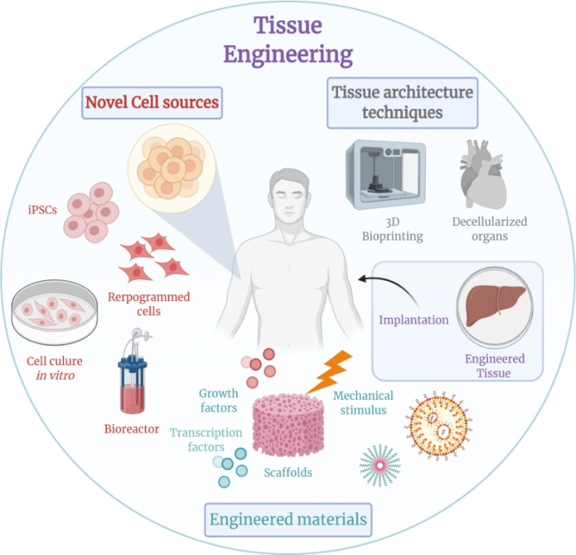
One of the most fascinating multidisciplinary study areas that combines engineering and life science principles is tissue engineering [25]. It primarily entails the utilisation of living cells and the creation of biological replacements that can be inserted into a site of tissue damage for repair. A key component of tissue engineering stems from the creation of polymeric scaffolds, which allow for a strong connection between seeded cells and newly formed organs. A scaffold needs to meet a few fundamental qualities in order to be a successful temporary ECM. The scaffold must, above all, be biocompatible and encourage cell multiplication while preventing an immunological response. Second, nutrition transport, cellular ingrowth, and vascularization all depend on a linked, highly porous 3D microenvironment. Thirdly, because the scaffolds are often made of biodegradable polymers, the pace of degradation should be planned to match or at least be comparable to the rate of tissue regeneration. The porous scaffold must also have enough mechanical qualities to preserve its structural integrity and avoid collapsing. Over the past few decades, there has been a lot of research on tissue-engineering scaffolds, and as a result, many methods have been proposed to help with tissue regeneration. These methods include solvent casting and particle leaching, molecular self-assembly, thermally induced phase separation, electrospinning, and others. Among these, electrospinning has grown in favour among tissue engineers because it offers a flexible and affordable way to make scaffolds with intricate biomimetic structures. Additionally, a wide variety of polymers, including synthetic, wholly natural, composite mixes, and even organ-specific extracts, can be used to fabricate electrospun fibres. What’s more intriguing is that by adjusting the manufacturing conditions or the polymer compositions, the mechanical and biological characteristics of the electrospun scaffolds may be easily controlled. These benefits have led to the use of electrospun techniques for the engineering of different skeletal muscle, bone, cartilage, skin, blood vessel, and neural tissues. It is important to note that, in contrast to the proteolytical degradation method used by matrix metalloproteinases to break down natural ECM, practically all electrospun scaffolds are either non-degradable or break down hydrolytically. Recent research has led to the development of a reactive macromer with fluorescent and protease-cleavable peptides that can be photo-polymerized to generate an electrospun fibrous hydrogel. A novel biomimetic method to produce protease-sensitive fibrous scaffolds, this biomimetic scaffold is vulnerable to protease-mediated cleavage in vitro and may monitor degradation in vivo utilising transdermal fluorescence imaging in a subcutaneous mouse model. Non-invasive tracking of biomaterials is essential to assess the pace of implant breakdown in vivo and demonstrate its function in tissue regeneration. To enable tracing, current biomaterials must always be labelled with fluorescent dyes or nanoparticles. A highly porous artificial extracellular matrix is required for tissue engineering in order to sustain and direct cell development and tissue regeneration. Such scaffolds have been made using biodegradable polymers, both natural and manmade. In order to replicate the extracellular matrix found naturally in bones, nanofiber scaffolds are employed in bone tissue engineering. The organised structures that make up the bone tissue are either arranged in a compact or trabecular form and range in length from a few centimetres to just a few nanometers. The nanocomposite structure of the bone ECM is made up of non-mineralized organic components, mineralized inorganic components, and numerous other non-collagenous matrix proteins. The ECM is flexible and strong thanks to the inorganic mineral salts and organic collagen fibres. Although the bone is a dynamic tissue capable of self-healing after minor wounds, it is unable to regenerate after suffering from big abnormalities like bone tumour resections and severe non-union fractures because it lacks the proper template. Currently, autografting is the go-to treatment, which entails taking donor bone from a small, easily accessible area of the patient’s own body and transplanting it into the damaged area.
Due to its reliable integration with the host bone and ability to prevent immune system problems, autologous bone transplantation offers the greatest clinical success [26]. However, because of its limited availability and donor site morbidity brought on by the harvest process, its use is restricted. Additionally, because autografted bones lack blood vessels and must obtain nutrients by diffusion, their survival in the host is compromised. Due to the body’s fast rate of remodelling, the grafts may also be reabsorbed before osteogenesis is finished. Allografting, which involves transplanting bones taken from a human corpse, is another treatment option for addressing severe bone deterioration. Allografts, however, increase the host’s chance of contracting illness and infection. A flexible solution to treat bone fractures and deformations is provided by bone tissue engineering. The design and properties of the natural extracellular matrix are particularly well mimicked by nanofibers created using electrospinning. Biologically active substances that encourage tissue regeneration can be delivered using these scaffolds. In a perfect world, these bioactive substances would be osteoinductive, osteoconductive, and osseointegratable. Bioactive ceramics, bioactive glasses, and synthetic and biological polymers are examples of bone substitute materials that are used to replace autologous or allogeneic bone. The idea behind bone tissue engineering is that over time, the body’s own freshly regenerated biological tissue will reabsorb the materials and replace them. Bone tissue engineering is not the only use of tissue engineering; significant research is also being done in the fields of cartilage, ligaments, skeletal muscle, skin, blood vessels, and brain tissue engineering.
1.4.2 Drug Delivery System
The selection of the drug carrier is crucial for the effective delivery of medicines to the intended target [27]. Maximum therapeutic effect after drug delivery to the target organ, circumvention of the body’s immune system while drug is in transit to the organ, retention of therapeutic molecules from drug preparation to final delivery, and proper drug release are the requirements for the ideal drug carrier. As a potential medication carrier candidate, nanofibers are now being studied. Due to their biocompatibility and biodegradability, which prevent damage to host tissue and toxic buildup in the body, respectively, natural polymers like gelatin and alginate make for suitable manufacturing biomaterials for carrier nanofibers. Nanofibers have a large surface area-to-volume ratio as a result of their cylindrical form. The fibres have a high drug-loading capacity as a result, and they are capable of dispensing medicinal molecules over a sizable surface area. For spherical vesicles, the surface area to volume ratio can only be altered by shifting the radius, whereas nanofibers offer more degrees of freedom by altering both the length and the cross-sectional radius. Their application in drug delivery systems, where the functioning parameters must be precisely controlled, benefits from this flexibility. According to preliminary research, antibiotics and anticancer medications can be enclosed in electrospun nanofibers by first dissolving them in the polymer solution. In order to prevent internal organs and tissues from adhering together after surgery, surface-loaded nanofiber scaffolds are helpful. Adhesion develops throughout the healing process and can result in issues like chronic pain and reoperation failure.
Several anatomical pathways can be used to administer drugs to a human body. They could be aimed at certain organs and disorders, or they could be designed to have systemic effects. Depending on the disease, the desired outcome, and the product in stock, the route of administration is chosen. Drugs can either be injected into the area of the body that is diseased or they can be injected systemically and specifically into the area that is diseased. The development of electrospun polymer nanofibers and their use in biomedical applications is being approached more broadly, with a key focus on drug delivery systems. As electrospun nanofibers have the potential to revolutionise the healthcare industry, these systems are essential. With their high surface area-to-volume ratio and adaptable qualities, electrospun polymer nanofibers make an excellent platform for regulated and targeted drug administration. These systems can offer sustained release patterns, enhance medication stability, and increase drug bioavailability by integrating therapeutic chemicals within the nanofibers or utilising them as carriers. Additionally, the adaptability of electrospun nanofibers offers customised drug delivery techniques for certain purposes, including cancer therapy, tissue regeneration, and wound healing. The use of electrospun nanofibers in drug delivery systems holds promise for more effective and patient-centric treatment modalities, ultimately leading to improved healthcare outcomes, as the field continues to evolve. The development of electrospun polymer nanofibers for biomedical applications has increasingly incorporated drug delivery systems, encouraging new therapeutic approaches. A flexible platform for drug encapsulation and release, electrospun nanofibers enable fine control over drug dose and kinetics. Optimising drug distribution to target tissues, lowering systemic side effects, and sustaining therapeutic drug levels for extended periods of time are only a few of the problems that this capacity is particularly useful for. The advantage of electrospun nanofiber-based drug delivery systems is their capacity to meet the particular requirements of many biomedical applications. For example, in tissue engineering, nanofiber scaffolds can act as transporters for cytokines or growth factors, encouraging cell division and proliferation. These scaffolds can be made to release antimicrobial medicines or to encourage angiogenesis in wound healing, speeding up the healing process. Additionally, to improve their drug loading and release capabilities, electrospun nanofibers can be functionalized with different chemical groups, nanoparticles, or biomolecules. This adaptability creates possibilities for targeted drug delivery, where nanofibers can be made to release medications primarily where they are needed, reducing side effects that aren’t intended and enhancing therapeutic results. There is increased interest in fusing the advantages of electrospun nanofibers with personalised medicine strategies as the field of nanomedicine develops. Drug delivery methods could revolutionise treatment effectiveness while reducing side effects if they are tailored to specific patient profiles, genetic variables, and disease characteristics.
Modern medicine is not complete without drug delivery systems, which provide methods for enhancing pharmacological therapy for better patient results. These systems are designed to facilitate the administration, absorption, distribution, and release of medications throughout the body. In order to modulate pharmacokinetics, reduce side effects, increase bioavailability, and encourage patient compliance, their fundamental tenets centre on controlled and targeted drug release. The drug substance, delivery vehicle/matrix, release mechanism, route of administration, and control systems in more complex circumstances are all necessary parts of a conventional drug delivery system. These systems are used in a variety of administration methods, such as oral, injectable, transdermal, inhalational, and implantable approaches, as well as in specialised drug delivery techniques. The safe, effective, and patient-friendly administration of therapeutic drugs to their targeted locations is fundamentally dependent on drug delivery systems. Drug delivery systems are crucial components of contemporary medicine and are set to revolutionise the way therapeutic drugs are administered. Their main goal is to perfect and optimise the complete range of medication administration processes, from intake to distribution and finally release in the body. In addition to modifying pharmacokinetics, decreasing side effects, boosting bioavailability, and encouraging improved patient adherence to treatment regimens, these systems follow fundamental principles of controlled and precisely targeted drug administration. The therapeutic drug substance itself, the adaptable delivery vehicle or matrix, the mechanisms orchestrating drug release, the selected route of administration, and in more sophisticated iterations, sophisticated control systems incorporating real-time physiological feedback, are all essential components of a standard drug delivery system. The broad range of uses includes oral, injectable, transdermal, inhalational, and implantable techniques, each of which addresses certain medical conditions. Additionally, the development of tailored drug delivery devices enables precise drug placement, minimising collateral injury to healthy tissues while maximising therapy effectiveness. Overall, drug delivery technologies bring in a new era of healthcare by guaranteeing that medicinal substances get to their destinations quickly, safely, and with the patient’s best interests in mind.
1.4.3 Wound Dressing and Scaffolds
When it comes to supporting tissue healing and regeneration, wound dressings and scaffolds play crucial roles in the fields of tissue engineering and wound care [28]. By fostering the ideal environment for healing while guarding against pollutants and infections, wound dressings play a crucial part in wound management. They come in a variety of shapes and sizes, from conventional gauze dressings to cutting-edge hydrocolloid, foam, and alginate dressings. These dressings keep the wound moist, control exudate, and may even include bioactive ingredients to speed up the healing process. Scaffolds, on the other hand, act as structural frameworks in tissue engineering, offering assistance and direction for cell adhesion, proliferation, and differentiation. Scaffolds are made from materials that are biocompatible and frequently biodegradable, allowing them to break down gradually while new tissue develops. They are crucial in the regeneration of numerous tissues, including bone, cartilage, skin, nerves, and cardiovascular structures because of their mechanical characteristics, porosity, and surface changes that are specifically designed to imitate the target tissue. In essence, wound dressings and scaffolds are cutting-edge approaches to both tissue engineering and wound care that present prospects for better patient outcomes and the regeneration of harmed or deteriorated tissues. In the fields of wound care and tissue engineering, wound dressings and scaffolds serve crucial roles in promoting the healing and regeneration of injured or compromised tissue. In wound care, dressings act as barriers of protection and provide a healing environment. They occur in a variety of shapes and sizes, ranging from conventional gauze to cutting-edge hydrocolloid, foam, and alginate dressings. These dressings can incorporate bioactive substances to quicken the healing process in addition to absorbing exudate and preventing infections. Additionally, they keep the environment around the wound wet, supporting vital biological processes like angiogenesis and tissue repair.
Scaffolds, on the other hand, serve as fundamental building blocks in tissue engineering, directing the formation and development of new tissue. Scaffolds, which are built from materials that are both biocompatible and frequently biodegradable, provide a framework for cellular adhesion, proliferation, and differentiation. The mechanical, porosity, and surface features of the target tissue are painstakingly replicated in these structures, which make them the best options for regenerating a variety of tissues, including bone, cartilage, skin, nerves, and even cardiovascular components. The gradual replacement of degrading scaffolds over time by freshly generated tissue allows for a smooth transition from the scaffold to the patient’s own biological structure. Acute wound management and the complex process of tissue regeneration are both addressed by wound dressings and scaffolds, which constitute fundamental improvements in healthcare. These developments improve patient outcomes and have the potential to revolutionise how we treat illnesses and injuries, ultimately leading to an improvement in the quality of life for people who require tissue repair or wound care.
1.4.3.1 Classification of Wound Dressing
Materials used as wound dressings serve to keep a wound safe. Additionally, they serve as a barrier against viruses [29]. In order to hasten the healing process and lower the risk of infections, an ideal wound dressing has good qualities including good application, biocompatibility, stability, and flexibility. It also ensures a good gas barrier and biodegradability. To stop bacterial invasion, wound dressings must also be able to manage wound exudates. There are two types of wound dressings: primary and secondary. While the secondary dressing is used to cover the original wound dressing, the primary wound dressing is applied directly to the area that was injured. Materials for wound dressings have a variety of uses. In order to categorise them, they are divided into four groups: traditional/passive dressings, skin substitutes, interactive/artificial dressings, and bioactive dressings. To stop bleeding and stop the wound from coming into contact with the environment again, traditional/passive wound dressings are typically applied during the first stage of treatment. These dressings have drawbacks, such as the potential for bleeding, low vapour permeability, and the potential to harm the newly formed epithelium after removal. Exudates from these dressings may leak and cause bacterial infections. The traditional dressing examples, which stand out for their great absorption ability, include tulle, gauze, and gauze cotton composites. Allografts, xenografts, and tissue derivatives are further terms for biological dressings known as skin substitutes. Allografts, which are pieces of fresh or frozen skin taken from donors, have a limited usage due to immunological reactions that cause the body to reject them. Allografts have a number of drawbacks, including the possibility of infection, the spread of disease, and their high cost and short shelf life. A xenograft is an organ or tissue transplant from a donor of a different species to the recipient. Wound dressings that are interactive or artificial are frequently made of biopolymers and synthetic polymers. Gelatin, alginate, chitosan, and other biopolymers are the most often utilised. The various types of artificial wound dressings include foams, films, composites, sprays, etc. The majority of them are transparent. Permeable to oxygen and water vapour yet impermeable to bacteria are polymeric films and foams. These bandages are appropriate for wounds with little exudate. The benefits of interactive wound dressings are their affordability, dependability, and longer shelf life. Bioactive substances, such as antimicrobials and growth factors, are incorporated in bioactive wound dressings made from biopolymers to speed up the healing of wounds. Collagens, alginate, hydrocolloids, and hydrofibres are a few examples of biopolymers.
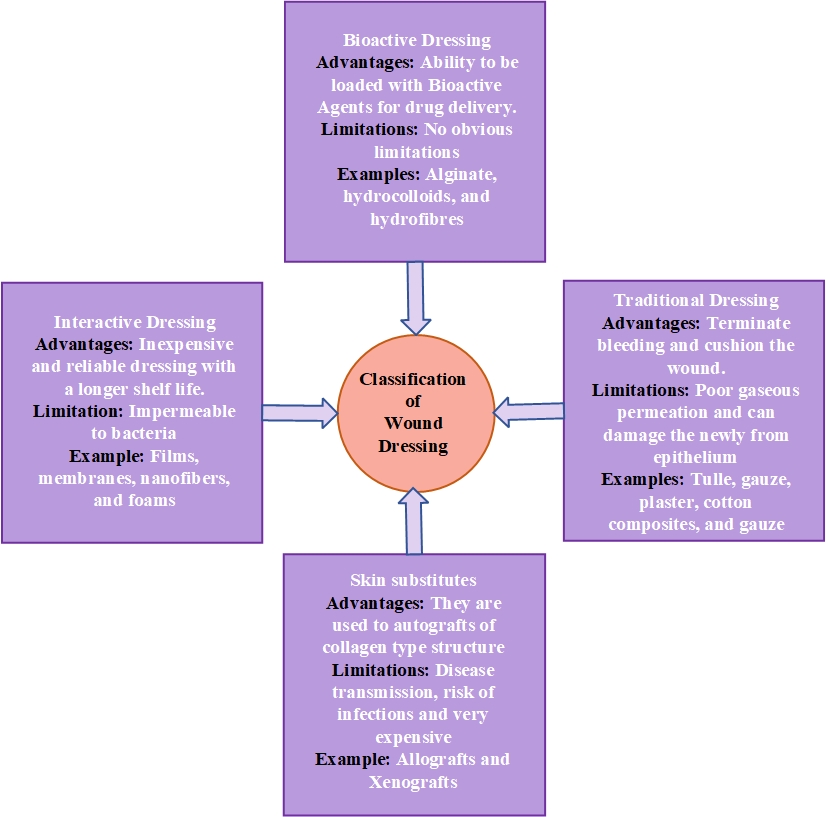
Skin substitutes are biological dressings and they are additionally classified as allografts, xenografts, and tissue derivatives. Allografts are fresh or freeze-dried skin fragments collected from donors and their use is limited by immune reactions, resulting in rejection by the body. Disadvantages of allografts include disease transmission, risk of infections and they are very expensive with limited shelf life. A xenograft is a tissue graft or organ transplant from the recipient from a donor of a different species. Interactive/artificial wound dressings are frequently formulated from synthetic polymers and biopolymers.
Gelatin, alginate, chitosan, and other biopolymers are the most often utilised. The various types of artificial wound dressings include foams, films, composites, sprays, etc. They are generally made of transparent polymeric films and foams that are impervious to bacteria but permeable to oxygen and water vapour. These bandages are appropriate for wounds with little exudate. The benefits of interactive wound dressings are their affordability, dependability, and longer shelf life. Bioactive substances, such as antimicrobials and growth factors, are incorporated in bioactive wound dressings made from biopolymers to speed up the healing of wounds. Collagens, alginate, hydrocolloids, and hydrofibers are a few examples of biopolymers.
1.4.4 Biosensing and Diagnostics
A variety of bioactive substances, including enzymes, antibodies, and molecular probes, can be included in nanofibers to give them the ability to interact with certain biological molecules. As a result of this interaction, precise and sensitive detection mechanisms are produced, which are essential for biosensing and diagnostics. The high surface area-to-volume ratio and programmable characteristics of nanofibers make them excellent substrates for biosensors because they improve molecule recognition and signal transmission. One of the significant uses is in the creation of electrochemical biosensors, where the addition of electroactive materials to nanofibers allows for the quick and precise detection of infections or biomarkers. Additionally, the larger density of biorecognition components can be immobilised on electrospun nanofibers, increasing sensitivity and lowering detection limits. Additionally, the real-time examination of clinical samples like blood, saliva, or urine is provided by these nanofiber-based biosensors that can be incorporated into portable and point-of-care diagnostic tools. By enabling early detection and monitoring of ailments ranging from infectious diseases to chronic illnesses, this technology has the potential to revolutionise disease diagnosis and have a huge impact on public health.
Additionally, wearable biosensors that can continually monitor physiological indicators have been developed thanks to the adaptability of electrospun nanofibers. These devices open the door to individualised healthcare and remote patient monitoring. A biosensor is an analytical tool that uses a transducer to convert molecular recognition of a target analyte into a quantifiable signal. The glucose sensor, which was introduced 30 years ago in its current form and has had a transformative impact on the care of diabetes, is the most well-known example currently in use. Examples of commonly used lateral flow assays include at-home pregnancy tests. Biosensors hold the promise of a user-friendly, sensitive, and affordable technology platform for infectious diseases that may quickly identify pathogens and foretell effective therapy. Small fluid volume manipulation has benefits such as quick test times, low energy usage, high mobility, high throughput, and multiplexing capabilities. Recent developments in micro- and nanotechnologies have produced biosensors that can carry out the intricate molecular assays necessary for many infectious illnesses. Parallel to this, a great deal of work has been done in understanding the genomes and proteomics of pathogens and how they interact with the host. Serology, which involves the multiplex detection of host immune response antibodies, may increase the overall specificity whereas biosensor-based immunoassays may increase the sensitivity of pathogen-specific antigen detection. Additional system integration might make it easier to build assays that incorporate targets specific to pathogens as well as indicators of host immune responses at various phases of infection.
1.5 Polymer Selection for Nanofiber Production
A key factor in determining the eventual effectiveness and usefulness of the nanofibers in a variety of applications, including but not limited to tissue engineering, drug administration, filtration, and wound healing, is the choice of polymer for their manufacture. The final nanofibers’ mechanical, chemical, and physical characteristics are greatly influenced by the polymer used. Researchers and engineers need to take into account details like the desirable biocompatibility, rate of degradation, mechanical strength, and hydrophilicity or hydrophobicity of the nanofibers in relation to the planned application. Polylactic acid (PLA), poly (lactic-co-glycolic acid) (PLGA), and polycaprolactone (PCL) are examples of biodegradable polymers that are frequently used in tissue engineering because they give structural support while slowly dissolving to enable tissue regeneration. On the other hand, polymers may be utilised for drug administration because of their capacity to encapsulate and release medications in a regulated manner. The flexibility of polymer choice is highlighted by its capacity to be tailored to fit particular objectives, enabling researchers to align nanofiber characteristics and behaviours with the needs of a wide range of applications. In essence, the precise engineering of nanofibers relies on the careful selection of polymers as a cornerstone, allowing these adaptable materials to be tailored to tackle certain biological, industrial, and environmental problems. The performance and application of the produced nanofibers are significantly influenced by the varied choice of polymer used in nanofiber manufacture. Choosing the polymer is similar to choosing the material that will serve as the base for the complete nanofiber structure. It depends on a variety of elements, including the required electrical conductivity, biocompatibility, degradation characteristics, and desired mechanical properties. Due to their capacity to offer transient structural support while gradually giving way to tissue regeneration or drug release, biodegradable polymers including PLA, PLGA, and PCL are frequently used for applications in tissue engineering and drug delivery. Additionally, the diameter, shape, and mechanical strength of the nanofiber are greatly influenced by the molecular weight and polymer concentration. To acquire the ideal nanofiber qualities, researchers must painstakingly adjust these parameters because even little changes can have a significant impact on the final product. Furthermore, polymer solvent interactions are crucial in defining the simplicity of electrospinning and the quality of the resulting nanofibers. So choosing the right solvent or solvent mixture is crucial to the electrospinning process. Furthermore, the choice of polymer can be adjusted to include additives or nanoparticles that confer these features when constructing nanofibers for particular purposes, such as wound dressings needing antimicrobial capabilities or filtration membranes requiring high porosity. Due to their adaptability, multifunctional nanofibers can be produced, extending their usefulness across a wide range of sectors.
1.6 Optimization of Electrospinning Process
The quality, uniformity, and application of the produced nanofibers are all influenced by the electrospinning process, which must be optimised for best results. This complex procedure requires the systematic fine-tuning of a number of variables and conditions, each of which is essential in determining the morphology, diameter, alignment, and overall performance of the nanofiber. The features of the final product are influenced by a wide range of variables, including the polymer content, solvent selection, applied voltage, flow velocity, and spinneret-collector distance. It is crucial to choose the right polymer type, molecular weight, and solvent mix to produce nanofibers that meet the demands of the desired application, including those related to biocompatibility, mechanical strength, and degradation rates. The electrospinning arrangement must also be customised to the particular goals and materials being used, taking into account spinneret design, collector type, and environmental factors. The nanofibers are further refined via post-processing procedures like annealing, cross-linking, or surface modification, which improve their mechanical, chemical, or biological properties. When moving from laboratory-scale to industrial-scale manufacturing, the optimisation process also includes cost-effectiveness, sustainability, and scalability factors. Adjustments and improvements are guided by continuous monitoring and characterization using methods like mechanical testing and scanning electron microscopy, guaranteeing that the electrospinning process produces nanofibers of the greatest quality and relevance to the intended applications. Overall, optimisation is essential to realising the full potential of electrospun polymer nanofibers and advancing the frontiers of nanotechnology and its revolutionary applications in a wide range of fields.
The development of electrospun polymer nanofibers at various scales and for a wide range of applications is influenced by the complex and diverse process of electrospinning optimisation, which extends beyond the confines of the laboratory. Each parameter in this procedure is thoroughly investigated because they can all have a big impact on the characteristics of the finished nanofibers. Researchers carefully choose the polymer kinds, molecular weights, and solvent compositions that correspond with the needs of the intended application as they deftly navigate the complexities of creating polymer solutions. In order to avoid problems like nozzle clogging during electrospinning, proper solvent handling is essential. The electrospinning apparatus itself can be improved. Nanofiber morphology and alignment are influenced by variables such as spinneret design, collector type, and environmental conditions (such as humidity and temperature). Whether the purpose of the experiment is to generate tissue engineering scaffolds, drug delivery vehicles, or filtration membranes, researchers must modify the setup to achieve their particular objectives. Techniques for post-processing are also essential to optimisation. These processes, which may involve surface modification, cross-linking, or annealing, fine-tune the properties of nanofibers to improve their mechanical strength, chemical reactivity, or biocompatibility. Additional difficulties with regard to production rate, quality assurance, and cost-effectiveness arise when the electrospinning process is scaled up from laboratory-scale to industrial-scale manufacturing. To ensure the practicality and financial viability of large-scale nanofiber production, optimisation efforts must take these considerations into account. Recent years have seen a rise in the importance of sustainability issues, leading researchers to optimise the electrospinning procedure with an eye towards reducing material waste, energy use, and environmental impact. These initiatives support the overarching objectives of environmentally responsible manufacturing. Throughout the optimisation process, ongoing characterization and monitoring are crucial. Researchers evaluate the shape, diameter, mechanical properties, and surface chemistry of nanofibers using cutting-edge methods like scanning electron microscopy (SEM), atomic force microscopy (AFM), and mechanical testing. These results guide on-going modifications and improvements, assisting scientists in obtaining the appropriate nanofiber characteristics and satisfying certain requirements for their intended applications.
1.7 Characterization of Electrospun Nanofiber
1.7.1 Morphology Analysis
Analysing the physical characteristics of electrospun nanofibers is one of the key parts of characterisation. Nanofiber morphology, including their diameter, alignment, and porosity, can be seen using methods like scanning electron microscopy (SEM) and transmission electron microscopy (TEM). These findings are crucial for determining the overall standard and reliability of nanofiber production.
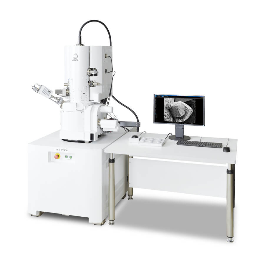
One of the main methods for morphologically analysing nanofibers is scanning electron microscopy (SEM). To produce high-resolution photographs, it entails blasting the nanofiber surface with electrons and gathering the secondary electrons that arise [30]. Researchers can see the morphology of nanofibers at the micro- and nanoscale using SEM, which provides details on things like fibre diameter, surface roughness, and alignment. Researchers can evaluate the consistency and calibre of nanofiber production by looking at SEM pictures. In the field of nanomaterials, materials science, and other scientific disciplines requiring high-resolution surface research, scanning electron microscopy (SEM) is a key imaging technique. [31]. SEM works by focusing an electron beam onto a specimen’s surface and then detecting the signals produced by the electrons’ interactions with the sample. These signals, which are mostly secondary electrons (SE) and backscattered electrons (BSE), offer priceless information on the specimen’s topography, morphology, and composition. SEM is a flexible technique for both qualitative and quantitative investigation since SE imaging is particularly good for showing surface details and microstructures and BSE imaging is sensitive to differences in elemental composition.
An electron source, electromagnetic lenses for beam control, a specimen stage for manipulating samples, detectors for signal collecting, and sophisticated control systems with imaging software are all crucial parts of SEM devices [32]. Emitting electrons from the source, concentrating and directing them towards the specimen, and evaluating the signals produced upon interaction with the sample are the steps in the procedure. Backscattered electrons, which are influenced by chemical composition, aid in compositional analysis, while secondary electrons, low-energy emissions linked to surface characteristics, provide fine topographical information. The broad range of SEM’s applications, including those in materials science, nanotechnology, biology, geology, forensics, quality assurance, environmental science, and archaeology, highlights the technology’s crucial role in illuminating and describing the micro- and nanoworld. SEM is continually developing, adding better imaging capabilities and extending its application to many fields of study and industry. Across a wide range of fields, scanning electron microscopy (SEM) is a revolutionary imaging method that probes deeply into the micro- and nanoscale world, revealing precise details of surface structures and elemental compositions. SEM is based on the basic idea of scanning a concentrated electron beam over a specimen’s surface, and it gathers vital data by seeing how the electrons interact with the sample. SEM devices have a complex range of parts that have been painstakingly engineered to produce accurate imaging and analysis. Electrons are emitted from the electron source, such as a tungsten filament or a field emission gun (FEG), and are later guided and focussed by electromagnetic lenses. While detectors catch the secondary electrons (SE) and backscattered electrons (BSE) produced during the electron-sample interaction, the specimen stage’s flexibility enables researchers to manipulate samples and see them from various angles. Important information about topography, morphology, and elemental distribution can be gleaned from these signals.
Beginning with the emission of electrons from the source and their precise focussing onto the sample, the operation of a SEM is a symphony of electron dynamics. SEs are released from the specimen’s surface as the electron beam interacts with it as a result of excitation, providing exquisite topographical information with sub-nanometer precision. BSEs, on the other hand, offer a window into compositional analysis due to the influence that atomic number variations have on their energy. SEM is extremely adaptable and has applications in a wide range of industries. It reveals the microstructure and surface properties of materials in materials research, assisting in the creation of novel materials. The capacity of SEM to visualise nanoparticles and nanoscale structures promotes nanotechnology, enabling ground-breaking research and innovation. By analysing cellular and tissue structures in biology, SEM opens up new perspectives, and in geology, it unlocks the secrets of minerals and rock formations. SEM is used in forensics to examine samples from crime scenes and track evidence, aiding in criminal investigations. SEM is used in manufacturing quality control to check components for flaws and ensure product integrity.
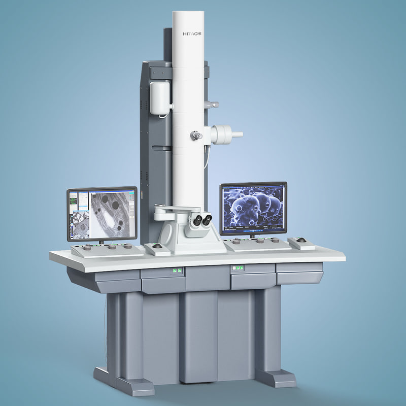
TEM is a sophisticated imaging method that provides resolution that is even higher than SEM. It works on the idea of sending electrons through incredibly thin nanofiber sections [33]. The interior structure of nanofibers, including their core-shell architecture, crystallinity, and nanoscale characteristics, may be thoroughly studied by TEM [34]. This method is very beneficial for a thorough examination of the composition and structure of nanofibers. In the field of nanoscale imaging, transmission electron microscopy (TEM) is regarded as an astonishing technological wonder. At its core, TEM works by passing a high-energy electron beam through a narrow specimen that has been painstakingly prepared, revealing the secret world of the nanoscale with unparalleled precision. The idea is deceptively straightforward yet incredibly effective: as electrons move through the sample, they interact intricately with its atoms to produce interactions like scattering, diffraction, and absorption. When these interactions are captured and recognised, a wealth of knowledge about the specimen’s innermost secrets is revealed.
Applications for TEM are as varied as the fields it supports. With atomic precision, it examines crystal structures, flaws, and microstructures in materials science, paving the way for improvements in material development. To characterise nanoparticles, nanomaterials, and complex nanodevices, and to create the foundation for ground-breaking breakthroughs, TEM is the virtuoso in the field of nanotechnology. It sheds light on the tiny realm of life by exposing the hidden landscapes of cells, tissues, and subcellular organelles in biology. To better comprehend Earth’s past, geologists use TEM to examine minerals, rocks, and geological objects at the nanoscale. Researchers in catalysis learn more about catalyst nanoparticles and their catalytic activities, which are essential for the creation of cleaner and more effective chemical reactions. Through the delivery of insights at the atomic and nanoscale levels that support innovation and advancement in a wide range of disciplines, TEM plays a crucial role in expanding the frontiers of human knowledge across a wide range of materials and scientific domains. Our understanding of matter at the atomic and molecular levels is constantly changing thanks to transmission electron microscopy (TEM), which continues to represent an unmatched frontier in the field of nanoscale inquiry [35]. TEM takes us on a voyage into the minute world of nanoparticles, crystalline structures, and biological subtleties by conveying a focussed stream of electrons through a specimen thinner than a human hair.
In order to produce a rich tapestry of data, TEM fundamentally relies on electron interactions with the object. These interactions, which also include inelastic scattering, which shows the specimen’s energy losses, and elastic scattering, which reveals surface morphology, result in intricate pictures and electron diffraction patterns that have the atomic arrangement of the material imprinted on them. The TEM equipment is an engineering marvel in and of itself. It consists of an electron source that emits high-energy electrons, magnetic lenses that precisely concentrate the beam, and detectors that catch the transmitted electrons to produce images and spectra. The specimen must endure the electron bombardment after being painstakingly prepared to be electron-transparent, which is a testimonial to the skill of TEM specimen preparation. As varied and extensive as the discoveries made possible by TEM, so are its applications. TEM sheds light on crystallography, flaws, and microstructures in the complex field of materials science, paving the way for new materials with specialised features. TEM is the alert eye that investigates the component parts of nanomaterials, enabling the development of cutting-edge technology. By revealing the inner workings of cellular organelles, biomolecules, and even viruses, TEM provides insights into the cellular world in the life sciences. Geologists use TEM to unravel the puzzles of minerals and rocks at scales previously imagined, providing insight into the planet’s geological past.
1.8 Summary
This field of study focuses on the development and usage of ultrafine polymer nanofibers for a variety of biomedical applications that are electrospun. These applications include biosensing, diagnostic instruments, medication delivery systems, tissue engineering, and wound healing. It takes careful optimisation of variables like polymer selection, concentration, solvent preference, and electrospinning conditions to create electrospun polymer nanofibers. The goal of this optimisation is to produce nanofibers with properties that are specifically suited to biomedical requirements, such as controlled drug release, mechanical strength, and biocompatibility. The incorporation of these nanofibers into sensors and devices benefits biosensing and diagnostic tools by enabling precise and sensitive detection of biomarkers and infections. Real-time monitoring and early disease diagnosis are possible with this technology. The creation of electrospun polymer nanofibers and their use in biomedicine represent a cutting-edge, multidisciplinary area with enormous promise to promote regenerative medicine, advance healthcare, and improve disease detection and treatment. It highlights the crucial role of nanotechnology in tackling important biomedical problems and opening the door for novel healthcare solutions.
References
- Farias, B.V., Pirzada, T., Mathew, R., Sit, T.L., Opperman, C. and Khan, S.A., 2019. Electrospun polymer nanofibers as seed coatings for crop protection. ACS Sustainable Chemistry & Engineering, 7(24), pp.19848-19856.
- Cui, J., Li, F., Wang, Y., Zhang, Q., Ma, W. and Huang, C., 2020. Electrospun nanofiber membranes for wastewater treatment applications. Separation and Purification Technology, 250, p.117116.
- Li, D., Yue, G., Li, S., Liu, J., Li, H., Gao, Y., Liu, J., Hou, L., Liu, X., Cui, Z. and Wang, N., 2022. Fabrication and applications of multi-fluidic electrospinning multi-structure hollow and core–shell nanofibers. Engineering, 13, pp.116-127.
- Wu, Y., Liu, Z., Wu, H., Zhang, K. and Liu, Q., 2023. Electrospinning Evolution Derived from TRIZ Theory for Directly Writing Patterned Nanofibers. Polymers, 15(14), p.3091.
- Eichholz, K.F., Gonçalves, I., Barceló, X., Federici, A.S., Hoey, D.A. and Kelly, D.J., 2022. How to design, develop and build a fully-integrated melt electrowriting 3D printer. Additive Manufacturing, 58, p.102998.
- Eslahi, N., Lotfi, R., Zandi, N., Mazaheri, M., Soleimani, F. and Simchi, A., 2022. Graphene-based polymer nanocomposites in biomedical applications. In Innovations in graphene-based polymer composites (pp. 199-245). Woodhead Publishing.
- Yadav, T.C., Srivastava, A.K., Mishra, P., Singh, D., Raghuwanshi, N., Singh, N.K., Singh, A.K., Tiwari, S.K., Prasad, R. and Pruthi, V., 2019. Electrospinning: an efficient biopolymer-based micro-and nanofibers fabrication technique. In Next Generation Biomanufacturing Technologies (pp. 209-241). American Chemical Society.
- Wang, X.X., Yu, G.F., Zhang, J., Yu, M., Ramakrishna, S. and Long, Y.Z., 2021. Conductive polymer ultrafine fibers via electrospinning: Preparation, physical properties and applications. Progress in Materials Science, 115, p.100704.
- Chakrapani, G., Ramakrishna, S. and Zare, M., 2023. Functionalization of electrospun nanofiber for biomedical application. Journal of Applied Polymer Science, p.e53906.
- Kachwal, V. and Tan, J.C., 2023. Stimuli‐Responsive Electrospun Fluorescent Fibers Augmented with Aggregation‐Induced Emission (AIE) for Smart Applications. Advanced Science, 10(1), p.2204848.
- Gera, S., Kankuri, E. and Kogermann, K., 2022. Antimicrobial peptides–unleashing their therapeutic potential using nanotechnology. Pharmacology & Therapeutics, 232, p.107990.
- Huang, C.Y. and Chiu, C.W., 2021. Facile fabrication of a stretchable and flexible nanofiber carbon film-sensing electrode by electrospinning and its application in smart clothing for ECG and EMG monitoring. ACS Applied Electronic Materials, 3(2), pp.676-686.
- Vijayakumar, S., Vijayalakshmi, K., Sangeetha, V. and Radha, E., 2023. Electrospinning—Material, Techniques and Biomedical Applications. In Nanobiomaterials (pp. 44-57). CRC Press.
- Wang, X. and Nakane, K., 2020. Preparation of polymeric nanofibers via immersion electrospinning. European Polymer Journal, 134, p.109837.
- Rajaram, R., Angaiah, S. and Lee, Y.R., 2023. Polymer supported electrospun nanofibers with supramolecular materials for biological applications–a review. International Journal of Polymeric Materials and Polymeric Biomaterials, 72(13), pp.1042-1058.
- Ahmadian, A., Shafiee, A., Aliahmad, N. and Agarwal, M., 2021. Overview of nano-fiber mats fabrication via electrospinning and morphology analysis. Textiles, 1(2), pp.206-226.
- Fan, Y., Tian, X., Zheng, L., Jin, X., Zhang, Q., Xu, S., Liu, P., Yang, N., Bai, H. and Wang, H., 2021. Yeast encapsulation in nanofiber via electrospinning: Shape transformation, cell activity and immobilized efficiency. Materials Science and Engineering: C, 120, p.111747.
- Jain, R., Shetty, S. and Yadav, K.S., 2020. Unfolding the electrospinning potential of biopolymers for preparation of nanofibers. Journal of Drug Delivery Science and Technology, 57, p.101604.
- Matysiak, W., Tański, T. and Smok, W., 2019. Electrospinning as a versatile method of composite thin films fabrication for selected applications. Solid State Phenomena, 293, pp.35-49.
- Barhoum, A., Pal, K., Rahier, H., Uludag, H., Kim, I.S. and Bechelany, M., 2019. Nanofibers as new-generation materials: From spinning and nano-spinning fabrication techniques to emerging applications. Applied Materials Today, 17, pp.1-35.
- Dubey, R., Dutta, D., Sarkar, A. and Chattopadhyay, P., 2021. Functionalized carbon nanotubes: Synthesis, properties and applications in water purification, drug delivery, and material and biomedical sciences. Nanoscale Advances, 3(20), pp.5722-5744.
- Waqas, M., Keirouz, A., Putri, M.K.S., Fazal, F., Sanchez, F.J.D., Ray, D., Koutsos, V. and Radacsi, N., 2021. Design and development of a nozzle-free electrospinning device for the high-throughput production of biomaterial nanofibers. Medical Engineering & Physics, 92, pp.80-87.
- Topuz, F., Abdulhamid, M.A., Holtzl, T. and Szekely, G., 2021. Nanofiber engineering of microporous polyimides through electrospinning: Influence of electrospinning parameters and salt addition. Materials & Design, 198, p.109280.
- Amini, S., Salehi, H., Setayeshmehr, M. and Ghorbani, M., 2021. Natural and synthetic polymeric scaffolds used in peripheral nerve tissue engineering: Advantages and disadvantages. Polymers for Advanced Technologies, 32(6), pp.2267-2289.
- Miranda, C.S., Ribeiro, A.R., Homem, N.C. and Felgueiras, H.P., 2020. Spun biotextiles in tissue engineering and biomolecules delivery systems. Antibiotics, 9(4), p.174.
- Nadine, S., Correia, C.R. and Mano, J.F., 2022. Engineering immunomodulatory hydrogels and cell-laden systems towards bone regeneration. Biomaterials Advances, p.213058.
- Ejazi, S.A., Louisthelmy, R. and Maisel, K., 2023. Mechanisms of Nanoparticle Transport across Intestinal Tissue: An Oral Delivery Perspective. ACS nano, 17(14), pp.13044-13061.
- Solarte David, V.A., Güiza-Argüello, V.R., Arango-Rodríguez, M.L., Sossa, C.L. and Becerra-Bayona, S.M., 2022. Decellularized tissues for wound healing: Towards closing the gap between scaffold design and effective extracellular matrix remodeling. Frontiers in Bioengineering and Biotechnology, 10, p.821852.
- Fan, C., Xu, Q., Hao, R., Wang, C., Que, Y., Chen, Y., Yang, C. and Chang, J., 2022. Multi-functional wound dressings based on silicate bioactive materials. Biomaterials, 287, p.121652.
- Bayrak, E., 2022. Nanofibers: Production, Characterization, and Tissue Engineering Applications. 21st Century Nanostructured Materials—Physics, Chemistry, Classification, and Emerging Applications in Industry, Biomedicine, and Agriculture, p.265.
- Shilpa Chakra, C., Divya, V., Shireesha, K., Madhuri, S., Rakesh Kumar, T., Uday Krishna, A. and Rakesh, D., 2022. Synthesis and Characterization of Emerging Nanomaterials. In Emerging Materials: Design, Characterization and Applications (pp. 37-102). Singapore: Springer Nature Singapore.
- Milani, M., Curia, R., Shevlyagina, N.V. and Tatti, F., 2023. Electron Microscopy Microscope Electron. In Bacterial Degradation of Organic and Inorganic Materials: Staphylococcus aureus Meets the Nanoworld (pp. 21-38). Cham: Springer International Publishing.
- He, B., Zhang, Y., Liu, X. and Chen, L., 2020. In‐situ transmission electron microscope techniques for heterogeneous catalysis. ChemCatChem, 12(7), pp.1853-1872.
- Morel, A., Oberle, S.C., Ulrich, S., Yazgan, G., Spano, F., Ferguson, S.J., Fortunato, G. and Rossi, R.M., 2019. Revealing non-crystalline polymer superstructures within electrospun fibers through solvent-induced phase rearrangements. Nanoscale, 11(36), pp.16788-16800.
- Zhang, Z., Cui, Y., Vila, R., Li, Y., Zhang, W., Zhou, W., Chiu, W. and Cui, Y., 2021. Cryogenic electron microscopy for energy materials. Accounts of Chemical Research, 54(18), pp.3505-3517.
Cite This Work
To export a reference to this article please select a referencing stye below:

Academic Master Education Team is a group of academic editors and subject specialists responsible for producing structured, research-backed essays across multiple disciplines. Each article is developed following Academic Master’s Editorial Policy and supported by credible academic references. The team ensures clarity, citation accuracy, and adherence to ethical academic writing standards
Content reviewed under Academic Master Editorial Policy.





