
Question 1 – What patient identifiers are present in the case scenario, and what specimen acceptance criteria should be followed?
Accurate identification of patients and precise specimen acceptance standards play critical roles in producing reliable laboratory findings and protecting patient safety in a complex healthcare environment. In this paper, the specific patient data contained in the medical scenario and laboratory report are considered, along with the criteria required for accepting samples in a diagnostic environment. Although the clinical situation does not identify a specific individual, it provides a detailed context from which demographic and medical history information can be derived (Allen and Cameron, 2017). Two sources of identifying information also arise within the laboratory report. First, the clinical scenario repeated in the report contributes to understanding the patient’s identity. Second, the information selected for reflection may reveal sensitive details such as the diagnosis and treatment plans, capturing important aspects of the individual’s medical experience.
Considering the case scenario of the 57-year-old female, the medical issues, chances of recovery, and effectiveness of treatment are important factors that can be influenced by patient identifiers such as age. Based on the provided case scenario, the patient’s date of birth, demographic details, and specimen information help guarantee accurate identification and linkage of clinical data. The patient’s age is helpful when choosing suitable treatment options. The specimen obtained from the left breast provides clinical detail that helps localize the mass. Proper observation and reporting, including the diagnosis of invasive ductal carcinoma, depend on these identifiers. Moreover, hormone receptor and HER2 status can influence personalized treatment. Tissue sections prepared using a microtome may also be used for further histological and immunohistochemical testing. These factors support accurate diagnosis, treatment planning, and effective communication among healthcare providers involved in the multidisciplinary management of breast cancer.
Turning to the specimen acceptance criteria, a strict framework is required to ensure the integrity and accuracy of samples and to support precise diagnostic outcomes. The first and most important condition is appropriate identification, which requires specimens to be properly labeled with a minimum of two distinct patient identifiers, such as the patient’s complete name, date of birth, health record number, or laboratory admission number (Evans et al., 2022). The consistency of these identifiers throughout the accompanying paperwork is critical in creating a dependable link to the patient’s information.
Appropriate management and collection are also required, which means following established procedures suitable for the specific tests. This includes using the correct collection containers, preservatives, and transport conditions. Any deviation from these guidelines requires proper documentation and communication with the laboratory, encouraging accountability. Timeliness is another important criterion in specimen acceptance. As stated by O’Grady and Morgan (2018), specimens should be delivered to the laboratory within the required time frame to prevent degradation that might compromise the accuracy of test findings. An acceptable volume and amount are also important so that the specimen is sufficient to complete the intended tests and to avoid inaccurate conclusions caused by an insufficient sample.
Question 2 – Why is clinical information important in histopathology, and how does it support diagnosis and treatment?
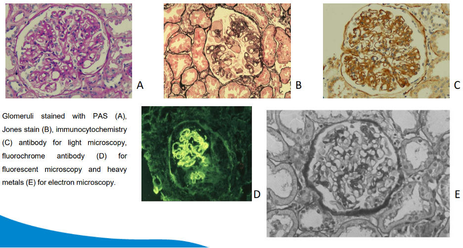
In histopathology, clinical information is essential for guiding accurate conclusions. In this case, understanding the characteristics of the cancer is supported by information about the poorly characterized mass, its size, location, and multifocality in the left breast. This is supported by gross evaluation and the presence of fibrofatty tissue. Microscopic examination further demonstrates grade 2 invasive ductal carcinoma with central high-grade DCIS (Dettmeyer, 2018). Clinical details guide the subsequent assessment of hormone receptors and can affect treatment choices. Histological staining approaches such as hematoxylin and eosin (H&E) are helpful in recognizing cell and tissue structures, while additional methods such as immunohistochemistry provide information about receptor status. This coordinated approach provides a more complete understanding of the pathology and supports personalized patient management. Clinical microscopes and microtomes are commonly used to process and analyze tissue samples from patients.
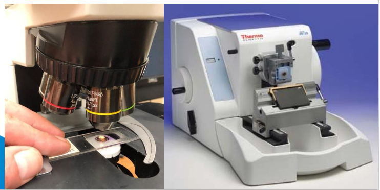
Furthermore, clinical information is critical for improving diagnostic accuracy. By providing context for histopathological results, clinical data allow pathologists to evaluate microscopic findings within the patient’s overall clinical condition. This contextual information is important in identifying small abnormalities that might otherwise go unnoticed, contributing to the accuracy of the diagnostic process. Beyond diagnosis, clinical information is important for prognosis and treatment planning. Although histopathology results are important, they may not always provide a complete picture of the patient’s condition or fully guide treatment recommendations (Allen and Cameron, 2017). When combined with clinical data, histopathology provides a more complete understanding of the illness and its possible course. This broader view supports the development of personalized treatment plans adapted to the needs of individual patients, promoting effective and patient-centered healthcare (Evans et al., 2022).
The incorporation of clinical information also improves communication and cooperation within the medical team. By sharing relevant patient data, pathologists, doctors, and other medical professionals can collaborate more effectively on treatment. Interprofessional collaboration supports a systematic and comprehensive approach to patient care and can contribute to better outcomes. Clinical data from histopathological cases not only have immediate therapeutic value but can also support continued research and improvement. Examining such cases may contribute to the development of new therapeutic strategies, preventive measures, and diagnostic tools that benefit medicine as a whole.
Question 3 – What is the importance of screening programs in pathology, particularly in relation to breast cancer?
Early diagnosis is made possible by screening programs, which support a healthcare approach in which preventive measures are given priority. These programs aim to detect precancerous changes or possible malignancies before symptoms appear so that treatment can begin promptly. Screening programs are important for early diagnosis, better outcomes, and reduced mortality in breast cancer pathology, which is relevant to the case scenario (Seely and Alhassan, 2018). Standard screening tests are designed to identify disease earlier, when treatment may be more effective.
In this case, the breast lesion might have been identified earlier through a screening program such as mammography. Considering the female patient in the scenario, the use of screening and early detection may be beneficial because it can identify suspicious changes before the condition becomes more advanced. Screening does not guarantee that every lesion will be detected before symptoms develop, but it can increase the opportunity for earlier diagnosis.
Screening initiatives also identify people who are at higher risk, allowing focused interventions and preventive measures. This approach can help reduce the number of patients affected by advanced chronic conditions through earlier detection and assessment of disease severity. Based on the principle that prevention is better than cure, early detection may also reduce the time and cost associated with later treatment. It can improve the patient’s chances of receiving timely care because diagnostic information is obtained earlier. A clearly defined target group based on demographics, risk factors, and other relevant criteria helps ensure that screening activities are directed toward the areas where they are most needed.
Quality Control. Standardized procedures for collecting specimens, carrying out tests, and interpreting results help maintain the quality of the screening process. Quality assurance is necessary to obtain accurate and reliable results.
System for Adequate Follow-up. To ensure that people with positive or abnormal screening results receive timely and appropriate medical attention, systems for proper follow-up are required.
Public Education. Educational initiatives can increase awareness of the benefits and limitations of screening programs. These initiatives encourage active participation and help individuals make informed decisions about their health. Examples of screening programs include Pap smears for cervical cancer, mammography for breast cancer detection, colonoscopy for colorectal cancer detection, and selected prostate-specific antigen (PSA) testing for prostate cancer. Academic Master’s discussion of population cancer screening programs provides further context on organized screening.
Question 4 – What do the patient’s ER, PR, and HER2 results indicate for treatment planning?
The consequences of this patient’s biopsy are important for her treatment plan, particularly in reducing the risk of disease progression and the spread of cancer cells. The positive estrogen receptor status with an Allred Score of 8/8 suggests responsiveness to hormone treatment and can guide the use of estrogen-blocking medication. The positive PR status may indicate a degree of responsiveness to hormone treatment and can also influence therapeutic decisions. HER2 status is essential because a HER2-negative result indicates that HER2 overexpression was not identified in this case, which means that HER2-targeted treatments such as trastuzumab would not be selected on the basis of HER2 positivity. This information helps clinicians develop an appropriate treatment strategy that may include surgery, chemotherapy, and hormone therapy while avoiding unnecessary interventions. The final diagnosis of invasive ductal carcinoma, combined with receptor status, supports a personalized approach intended to improve treatment effectiveness, reduce side effects, and improve the patient’s prognosis and quality of life.
Positive ER and PR values show that cancer cells are sensitive to hormonal influence. In other words, estrogen and progesterone can stimulate the tumor, so blocking these hormonal effects can slow cancer growth. Hormone treatments such as tamoxifen and aromatase inhibitors may help patients with ER-positive or PR-positive tumors. If the tumor is negative for both ER and PR, it is less likely to respond to hormone therapy. Alternative treatment options, including chemotherapy, may be considered in such circumstances.
HER2 is a receptor protein involved in cell growth. The HER2 protein is overexpressed in HER2-positive breast tumors, which can make them more aggressive. The HER2 result is therefore important when selecting the most appropriate treatment plan for the patient (Lamb et al., 2019).
If HER2-positive status is identified, targeted therapy may be required to improve treatment effectiveness and outcomes. In contrast, HER2-negative status may direct attention toward alternative treatment methods. Oncologists can develop a precise and individualized treatment approach after considering ER, PR, and HER2 status (Zhao and Gong, 2021). This highlights the importance of biological profiling in directing therapeutic options and reflects the wider development of personalized cancer care.
The incorporation of these biological markers into the patient’s treatment highlights the developing landscape of personalized medicine in cancer care.
Question 5 – What additional immunohistochemical, molecular, genetic, and imaging tests may be useful in this breast cancer case?
For the 57-year-old female patient, immunohistochemical (IHC) tests play an important role in breast cancer diagnosis and treatment decisions.
Ki-67 Immunohistochemistry (IHC). Ki-67 assesses the proliferation rate of cancer cells. Higher levels indicate more active cell division and can provide information about the aggressiveness of the tumor and its possible response to treatment.
p53 Mutation Examination. p53 IHC or genetic testing can identify abnormalities or mutations involving the p53 gene, which are often associated with more aggressive cancers. This information can contribute to treatment decisions when considered with the overall clinical and pathological findings (Botsikas et al., 2019).
MammaPrint Molecular Test. This genomic assay evaluates the expression of specific genes to estimate the risk of breast cancer recurrence. MammaPrint can help classify selected patients into lower- or higher-risk groups and can contribute to decisions about the need for adjuvant chemotherapy.
Breast cancer genetic testing examines the genetic landscape and may identify mutations that contribute to the development and progression of the disease. Testing for BRCA1 and BRCA2 mutations can identify genetic changes linked to an increased risk of breast and ovarian cancers, which may influence treatment and preventive decisions. Similarly, PIK3CA testing can identify mutations in a breast cancer-related gene and may contribute to treatment planning in selected cases. These developments are part of the broader use of genetic and molecular technologies in cancer care.
Immunohistochemistry is an important method for identifying and localizing particular proteins produced by cancer cells. Hormone therapy can be guided by ER and PR status, which indicate hormonal responsiveness. Assessment of HER2 expression helps determine whether HER2-targeted therapy is appropriate. Ki-67, which indicates the rate of tumor-cell proliferation, can also provide information relevant to prognosis and treatment choices.
In addition to these molecular findings, supplementary diagnostic tests can help provide a more complete clinical picture. MRI, or magnetic resonance imaging, provides detailed images that can be useful in selected situations, such as dense breast tissue or lesions that are difficult to evaluate with mammography (Kim et al., 2019). PET scans measure metabolic activity and may help with staging and monitoring treatment response. Blood tests are not diagnostic for breast cancer by themselves, but markers such as CA 15-3 may sometimes be used in monitoring the progression of established disease.
Reflection
Although I was thoroughly guided by the module leader, I initially faced challenges in identifying the issues mentioned in the case study. As a result, I was somewhat frustrated when I came across the first question related to patient identifiers. Because the patient identifiers were not listed separately, I found it difficult to select the most appropriate identifiers. Although explaining the importance of patient identifiers was relatively easy, choosing the most important factors was also challenging for me in this project.
Considering the next question, discussing the importance of clinical information was comparatively easier because I found most of the relevant information in the selected resources on breast cancer pathology. Although I understood the importance of screening programs in a general medical context, applying the importance of screening programs to the given case scenario was more difficult. I think answering the fourth question was the most difficult because it was directly related to the case study, and I had to explain the results of the specifically mentioned hormone receptors for the patient. This required additional time and effort related to clinical interpretation.
One of the major achievements I gained during this study was the opportunity to operate a microtome. Although clinical cutting tools are used for handling specimen pieces, a microtome is used for professional histopathological work such as preparing breast tissue sections for microscopic examination (Ajantha Devi and Nayyar, 2021). I observed that the specimen needed to be embedded properly in paraffin wax so that it remained stable during sectioning. This practical experience helped me understand the precision required when preparing tissue samples.
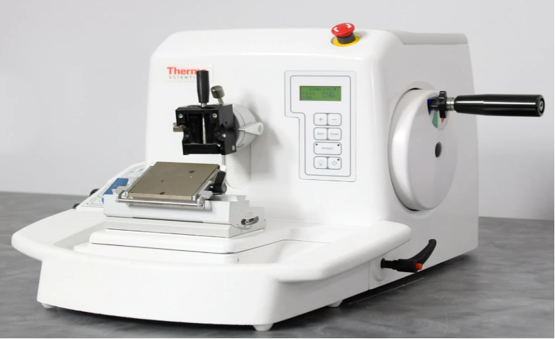
Based on my sustained efforts during this assignment, the activity went well overall. I was initially nervous about using the microscope and microtome, but my instructor’s assistance and clear directions reduced my concerns. Observing slides under the microscope was especially interesting, and using the microtome successfully gave me a sense of satisfaction. During the activity, safety was very important. I kept my fingers clear of the microtome blade and used gloves and safety glasses when working with chemicals. The instructor also repeatedly reminded us to avoid touching the eyes and face after processing tissue samples.
Throughout the exercise, my confidence in using the microscope and microtome increased significantly. Despite my early doubts, hands-on practice and the instructor’s guidance helped me become more confident. By the end, I felt comfortable using the microscope independently, preparing slides, and examining tissue samples. In future sessions, I would spend more time practicing how to draw microscopic features. Improving my drawing skills would allow me to represent observed features more accurately. In addition, I would actively seek more information about the individual tissue samples under investigation so that I could better understand their context and relevance. This additional knowledge would improve the overall learning experience.
References
Ajantha Devi, V. and Nayyar, A., 2021. Fusion of deep learning and image processing techniques for breast cancer diagnosis. Deep Learning for Cancer Diagnosis, pp.1–25.
Allen, D.C. and Cameron, R.I. eds., 2017. Histopathology Specimens: Clinical, Pathological and Laboratory Aspects. London: Springer London.
Botsikas, D., Bagetakos, I., Picarra, M., Da Cunha Afonso Barisits, A.C., Boudabbous, S., Montet, X., Lam, G.T., Mainta, I., Kalovidouri, A. and Becker, M., 2019. What is the diagnostic performance of 18-FDG-PET/MR compared to PET/CT for the N- and M-staging of breast cancer? European Radiology, 29, pp.1787–1798.
Dettmeyer, R.B., 2018. Forensic Histopathology: Fundamentals and Perspectives. Springer.
Evans, A.J., Brown, R.W., Bui, M.M., Chlipala, E.A., Lacchetti, C., Milner Jr, D.A., Pantanowitz, L., Parwani, A.V., Reid, K., Riben, M.W. and Reuter, V.E., 2022. Validating whole slide imaging systems for diagnostic purposes in pathology: guideline update from the College of American Pathologists in collaboration with the American Society for Clinical Pathology and the Association for Pathology Informatics. Archives of Pathology & Laboratory Medicine, 146(4), pp.440–450.
Galusha, H., 2021. What is a Microtome. Available at: https://newlifescientific.com/blogs/new-life-scientific-blog/what-is-a-microtome [Accessed 12 December 2023].
Kim, T.H., Yoon, H.J., Fouladdel, S., Wang, Y., Kozminsky, M., Burness, M.L., Paoletti, C., Zhao, L., Azizi, E., Wicha, M.S. and Nagrath, S., 2019. Characterizing circulating tumor cells isolated from metastatic breast cancer patients using graphene oxide based microfluidic assay. Advanced Biosystems, 3(2), p.1800278.
Lamb, C.A., Vanzulli, S. and Lanari, C.L.M., 2019. Hormone receptors in breast cancer: more than estrogen receptors.
O’Grady, S. and Morgan, M.P., 2018. Microcalcifications in breast cancer: From pathophysiology to diagnosis and prognosis. Biochimica et Biophysica Acta (BBA)-Reviews on Cancer, 1869(2), pp.310–320.
Seely, J.M. and Alhassan, T., 2018. Screening for breast cancer in 2018—what should we be doing today? Current Oncology, 25(s1), pp.115–124.
Zhao, H. and Gong, Y., 2021. The prognosis of single hormone receptor-positive breast cancer stratified by HER2 status. Frontiers in Oncology, 11, p.643956.
Cite This Work
To export a reference to this article please select a referencing stye below:

Academic Master Education Team is a group of academic editors and subject specialists responsible for producing structured, research-backed essays across multiple disciplines. Each article is developed following Academic Master’s Editorial Policy and supported by credible academic references. The team ensures clarity, citation accuracy, and adherence to ethical academic writing standards
Content reviewed under Academic Master Editorial Policy.





