
The skeletal system is a living organ system composed primarily of bones, cartilage, ligaments, marrow, and associated connective tissues. It provides structural support, protects vulnerable organs, stores important minerals, produces blood cells, and works with muscles and joints to create movement. The adult human skeleton is commonly described as having 206 bones, although the number can vary slightly because of accessory bones or differences in fusion, while children have more separate skeletal elements because many join together during growth. Bone itself is not an inert framework but metabolically active tissue that continually responds to mechanical loading, hormones, nutrition, injury, and age. Understanding the skeleton as living tissue explains why bone can grow, heal, remodel, weaken, and participate in mineral and blood-cell regulation throughout life. The skeletal system is therefore best understood not as a passive support structure but as a dynamic network whose tissues interact continuously with the muscular, endocrine, circulatory, immune, and nervous systems.
Organization of the Skeleton and Its Major Functions
The adult skeleton is divided into axial and appendicular components. The axial skeleton forms the body’s central framework and includes the skull, vertebral column, ribs, and sternum; in the standard adult count it contains 80 bones and is especially important for posture and protection of the brain, spinal cord, heart, and lungs. The appendicular skeleton contains 126 bones and includes the upper and lower limbs together with the pectoral and pelvic girdles. The upper limbs are adapted for mobility, reaching, grasping, and manipulation, while the pelvis and lower limbs provide weight bearing, balance, and locomotion. These divisions reflect both anatomy and function: the axial skeleton stabilizes and protects central structures, whereas the appendicular skeleton permits extensive movement through its connections with muscles and joints. Together they create a mechanical system in which bones act as levers, joints create points of controlled motion, and muscle contraction supplies force.
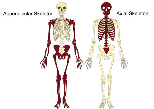
Figure 1. Axial and appendicular divisions of the human skeleton.
The skeleton also performs several physiological functions that are easy to overlook when attention is focused only on movement. Bone stores calcium and phosphate, minerals that are essential for nerve signaling, muscle contraction, blood clotting, and intracellular communication. Red marrow produces blood cells through hematopoiesis, while yellow marrow contains more adipose tissue and serves partly as an energy reserve. Cartilage provides flexible support and low-friction surfaces at many joints, ligaments connect bone to bone and stabilize articulations, and tendons usually connect muscle to bone so that muscular force can be transmitted to the skeleton. These tissues differ in composition, blood supply, healing capacity, and mechanical behavior, but they work together as an integrated musculoskeletal system. Bone also participates in endocrine signaling, which further demonstrates that the skeleton is biologically active rather than simply a rigid frame.
Long-Bone Structure, Compact Bone, and Spongy Bone
A typical long bone such as the femur contains a shaft called the diaphysis and enlarged ends called epiphyses, with the metaphysis positioned between them. During growth, the metaphysis contains the epiphyseal plate, a region of cartilage where lengthening occurs; after growth is complete, this plate is replaced by an epiphyseal line. The diaphysis surrounds a medullary cavity, while the epiphyses contain abundant spongy bone covered by a thinner layer of compact bone. Articular cartilage covers many joint surfaces and reduces friction while distributing load. The periosteum covers most outer bone surfaces except those covered by articular cartilage and contains vessels, nerves, connective tissue, and cells involved in growth and repair. The endosteum lines the medullary cavity and internal bone surfaces, helping support remodeling from within.
Compact, or cortical, bone forms the dense outer shell of bones and in many regions is organized into osteons, or Haversian systems, built from concentric lamellae surrounding central canals that contain blood vessels and nerves. Osteocytes occupy small spaces called lacunae and communicate through microscopic canaliculi, allowing cells embedded inside mineralized tissue to remain connected. Spongy, cancellous, or trabecular bone has a different architecture, consisting of a three-dimensional lattice of plates and struts with marrow and vessels in the spaces between them. Its lower density does not make it inherently weak. Trabeculae are arranged along patterns of mechanical stress and provide efficient strength with less mass than solid tissue. Diseases that thin or disconnect trabeculae can greatly reduce bone strength even when the external shape of the bone appears unchanged, which is one reason microarchitecture is clinically important.
Bone Matrix, Cells, Growth, and Remodeling
Bone matrix combines organic and mineral components whose mechanical properties complement one another. The organic portion is primarily type I collagen together with additional proteins, while the mineral portion consists largely of hydroxyapatite containing calcium and phosphate. Collagen provides tensile strength and flexibility, whereas mineral contributes hardness and resistance to compression. Healthy bone depends on the combination because tissue that is poorly mineralized can become soft and deformable, while abnormalities in collagen can make bone unusually fragile. Bone is maintained by several specialized cell types. Osteoblasts produce osteoid and help regulate mineralization, some later becoming osteocytes when surrounded by matrix. Osteocytes form an extensive communication network and help detect mechanical loading. Osteoclasts, large multinucleated cells derived from the monocyte-macrophage lineage, resorb bone so that old or damaged tissue can be removed and mineral balance maintained.
Bone modeling and remodeling explain how skeletal tissue changes across life. Modeling alters the size and shape of bone, particularly during growth, while remodeling replaces existing tissue through coordinated cycles of resorption and formation. Appropriate mechanical loading stimulates adaptation, whereas prolonged unloading can contribute to bone loss. Most bones develop through endochondral ossification, in which bone replaces a cartilage model, while intramembranous ossification forms bone directly within connective tissue and contributes especially to many flat bones of the skull and part of the clavicle. Longitudinal growth occurs at epiphyseal plates until they close after maturation, and appositional growth increases bone width. Genetics, growth hormone, thyroid hormone, sex hormones, nutrition, vitamin D, calcium, phosphate, physical activity, disease, and medication all influence these processes, demonstrating that skeletal development depends on both biological programming and environmental conditions.
Bone Shape, Joints, and Movement
Bones are commonly classified as long, short, flat, irregular, or sesamoid according to shape and mechanical function. Long bones such as the femur and humerus act as levers and contain recognizable shafts and ends. Short bones, including carpals and tarsals, provide stability with relatively limited movement. Flat bones such as the ribs, sternum, and many cranial bones protect organs and provide broad surfaces for muscle attachment, while vertebrae are classified as irregular because their complex forms do not fit other categories. Sesamoid bones develop within tendons where they can reduce friction, protect the tendon, or alter the direction of muscular force; the patella is the largest example. These shape categories are not arbitrary labels because each reflects the mechanical demands placed on a particular region of the skeleton.
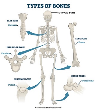
Figure 2. Major bone categories based on shape.
Joints, or articulations, connect bones and determine how movement occurs. Fibrous joints permit little or no movement and include skull sutures; cartilaginous joints unite bones through cartilage and allow limited motion, as in the pubic symphysis; and synovial joints contain a joint cavity, capsule, articular cartilage, and synovial fluid and permit the greatest range of movement. Hinge, pivot, ball-and-socket, saddle, condyloid, and plane joints are examples of synovial forms. Joint stability depends not only on bone shape but also on ligaments, muscles, tendons, and surrounding tissues. Greater mobility often involves a trade-off with stability, which helps explain why the shoulder moves through a wider range than the hip but is also more vulnerable to dislocation. Movement therefore depends on the coordinated properties of bone, cartilage, connective tissue, and muscle rather than on bone alone.
Mineral Homeostasis, Marrow, and Clinical Relevance
The skeleton acts as a regulated mineral reservoir rather than a passive storage room. Calcium is required for muscle contraction, nerve transmission, clotting, and cell signaling, so the body carefully regulates its movement between bone, blood, intestine, and kidneys. Parathyroid hormone, vitamin D metabolites, sex hormones, and local cellular signals influence absorption, excretion, bone formation, and bone resorption. Vitamin D supports intestinal calcium absorption and normal mineralization; severe deficiency can contribute to rickets in children and osteomalacia in adults. Bone marrow adds another major physiological role. Red marrow contains hematopoietic stem and progenitor cells that produce red blood cells, white blood cells, and platelets, while yellow marrow contains more fat but remains part of a biologically active microenvironment in which skeletal, vascular, and immune cells interact.
Clinical disorders demonstrate why skeletal biology must be understood in terms of living tissue. Fractures disrupt bone continuity and may also damage vessels, nerves, muscles, or joints, with healing proceeding through inflammation, callus formation, and later remodeling. Osteoporosis reduces bone strength through loss of mass and deterioration of microarchitecture, increasing fracture risk particularly at the hip, spine, and wrist. Osteomalacia differs because it primarily involves inadequate mineralization. Osteoarthritis affects the whole joint and prominently involves degeneration of articular cartilage, whereas rheumatoid arthritis is a systemic inflammatory autoimmune disease. Osteomyelitis is an infection of bone and requires a very different clinical approach. Adequate nutrition, weight-bearing and resistance exercise, avoidance of tobacco, fall prevention, and appropriate medical assessment all support skeletal health, but persistent pain, deformity, unexplained height loss, or serious injury requires professional evaluation because similar symptoms can arise from very different causes.
Conclusion
The skeletal system is a dynamic biological network rather than a static frame. Its axial and appendicular divisions provide support, protection, and movement; compact and spongy bone use different architectures to achieve mechanical strength; collagen and mineral give bone both flexibility and hardness; and osteoblasts, osteoclasts, and osteocytes continually build, remove, and monitor tissue. Growth, remodeling, joint structure, marrow, hormones, nutrition, and mechanical loading all contribute to skeletal function across the lifespan. Understanding bone as living tissue provides a stronger foundation for explaining development, fracture repair, osteoporosis, arthritis, mineral disorders, and lifelong skeletal health. The skeleton supports the body physically, but it also participates in blood formation, mineral regulation, and communication with other organ systems. Its strength therefore depends not on hardness alone but on a continuously regulated balance among structure, cells, circulation, movement, and metabolism.
References
National Institute of Arthritis and Musculoskeletal and Skin Diseases. “What Is Bone?”
Kamel-ElSayed, Suzan A., Trevor A. Nezwek, and Matthew A. Varacallo. “Physiology, Bone.” StatPearls, updated 10 Sept. 2024.
Tortora, Gerard J., and Bryan H. Derrickson. Principles of Anatomy and Physiology. Wiley, 2018.
Cite This Work
To export a reference to this article please select a referencing stye below:

Academic Master Education Team is a group of academic editors and subject specialists responsible for producing structured, research-backed essays across multiple disciplines. Each article is developed following Academic Master’s Editorial Policy and supported by credible academic references. The team ensures clarity, citation accuracy, and adherence to ethical academic writing standards
Content reviewed under Academic Master Editorial Policy.





