
Introduction
Polymerase chain reaction (PCR) is a laboratory technique for selectively amplifying a defined nucleic-acid sequence so that a target initially present in very small quantities becomes detectable and analyzable. The method transformed molecular biology because it allowed researchers to generate millions of copies of a chosen DNA region without first cloning that region in living cells. PCR is now routine in research laboratories, infectious-disease testing, inherited-disease analysis, oncology, forensic science, environmental monitoring, and sequencing workflows. Its basic cycle remains conceptually simple—denaturation, primer annealing, and extension—but reliable PCR depends on much more than repeating temperature changes. Primer design, template quality, enzyme choice, magnesium concentration, controls, contamination prevention, and interpretation all determine whether an apparent result represents the intended target. Modern thermal cyclers can complete reactions rapidly, and related methods such as reverse-transcription PCR, quantitative real-time PCR, multiplex PCR, and digital PCR have extended the same molecular logic to RNA, quantification, multiple targets, and absolute copy-number estimation (Ghannam & Varacallo, 2023; Khehra, Padda, & Zubair, 2025).
Molecular Components and the Thermal Cycle
A conventional PCR mixture contains template DNA, two sequence-specific primers, a thermostable DNA polymerase, deoxynucleotide triphosphates, magnesium ions, buffer, and water. The primers define the boundaries of the region to be amplified because one primer binds to each template strand on opposite sides of the target, with their 3′ ends oriented toward the sequence that will be copied. DNA polymerase cannot initiate synthesis without a primer, making primer placement a major determinant of specificity. Magnesium supports polymerase activity and influences primer-template binding; insufficient magnesium can reduce yield, while excessive magnesium can increase nonspecific amplification. Template DNA must contain the target and be sufficiently free from substances that inhibit the polymerase. A PCR assay therefore begins before the thermal cycler is started: sample collection, nucleic-acid extraction, primer design, reagent preparation, and controls all shape the result. Appropriate positive, negative, extraction, and internal controls are part of the analytical system rather than optional additions used only when a reaction fails.
The thermal cycle begins with denaturation, usually near 94–98°C, which separates double-stranded DNA into single strands by disrupting hydrogen bonding between complementary bases. The temperature is then lowered for annealing so that primers can bind to complementary sequences. Annealing temperature is chosen in relation to primer melting temperature and sequence composition rather than fixed at one universal value; temperatures that are too low encourage mismatched binding, while temperatures that are too high reduce primer binding and product yield. During extension, the polymerase adds nucleotides to the 3′ end of each primer, with Taq polymerase commonly working efficiently near 72°C. In an ideal early cycle, each target molecule produces two copies, and repeated cycling produces approximately exponential amplification until reagents become limiting and products begin to reanneal. Because reactions eventually plateau, conventional end-point product amount is not a simple linear measurement of the amount of starting DNA (Saiki et al., 1988).
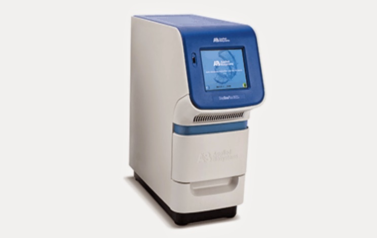

The images above illustrate PCR equipment and the concept of repeated thermal cycling, but an image or generic three-step description is not a complete experimental protocol. A reproducible assay must specify primer sequences, target region, reaction volume, polymerase system, reagent concentrations, cycling temperatures and durations, number of cycles, detection method, and acceptance criteria for controls. Primer pairs should also be examined for complementarity, secondary structure, repeated sequences, and unintended matches elsewhere in the genome. Modern hot-start enzymes can reduce nonspecific amplification before cycling begins, while high-fidelity polymerases provide proofreading activity when sequence accuracy is important for cloning or downstream sequencing. The historical development of PCR depended on several advances working together: oligonucleotide synthesis, understanding of DNA replication, thermostable polymerases such as Taq, automated thermal cyclers, and increasingly sensitive detection systems. Kary Mullis received a share of the 1993 Nobel Prize in Chemistry for the invention, but PCR became transformative because the surrounding chemistry and instrumentation made repeated amplification practical and scalable.
PCR Variants and Major Applications
Reverse-transcription PCR begins with RNA rather than DNA. Reverse transcriptase converts RNA into complementary DNA, which is then amplified by PCR, making the method useful for RNA viruses and studies of gene expression. Quantitative real-time PCR monitors fluorescence during amplification and uses the cycle at which signal becomes detectable to estimate starting target quantity, provided assay efficiency, controls, and calibration are appropriate. Multiplex PCR amplifies several targets in the same reaction and can conserve specimen and reagents, although primer compatibility becomes more complex. Digital PCR partitions a sample into many small reactions and estimates target concentration from the proportion of positive partitions using probability. These formats share the logic of sequence-specific amplification but answer different analytical questions. A conventional end-point PCR can answer whether a target product is detectable, qPCR can estimate relative or absolute quantity, RT-PCR enables RNA analysis, and digital PCR can support sensitive copy-number or low-frequency target measurement without relying on the same calibration approach used in standard quantitative PCR.
Applications span research, medicine, public health, and forensic science. Infectious-disease laboratories use PCR-based nucleic-acid amplification tests when culture is slow, difficult, or unsuitable. Genetic testing can amplify regions needed for variant analysis, carrier testing, or sequencing. Oncology laboratories may use validated PCR assays to identify tumor-associated variants or monitor measurable residual disease in selected conditions. Researchers use PCR to genotype organisms, confirm engineered constructs, clone genes, and quantify gene expression after reverse transcription. Forensic laboratories analyze defined genetic markers, while environmental scientists detect organisms through DNA recovered from water, soil, or other samples. Ancient-DNA work can amplify scarce historical material but requires unusually strict contamination control because tiny amounts of modern DNA can overwhelm the target. Across all these settings, PCR is powerful because it selectively amplifies sequence information, not because a positive amplification automatically proves disease, biological activity, or causation.
Quality Control, Limitations, and Interpretation
The same sensitivity that makes PCR useful also makes contamination a major risk. Trace amplified material from a previous reaction can generate a false-positive result if it reaches a new specimen. Laboratories therefore separate reagent preparation, specimen processing, and amplified-product analysis where appropriate and use no-template controls to detect contamination, positive controls to confirm reagent performance, extraction controls to monitor sample processing, and internal controls to detect inhibition. A negative result can arise because the target is truly absent, but it can also reflect poor specimen collection, low target concentration, degraded nucleic acid, inhibitors, primer-site variation, or testing at the wrong point in an infection or disease process. A positive result may detect residual nucleic acid from nonviable organisms rather than active infection. Interpretation must therefore integrate specimen quality, assay performance, symptoms, exposure, timing, and pretest probability rather than treating amplification alone as a complete clinical diagnosis (Khehra, Padda, & Zubair, 2025).
PCR also has technical limits in research. Standard Taq polymerase lacks proofreading activity and can introduce errors, making it less suitable when exact sequence fidelity is essential. Mixed samples can show amplification bias because some sequences amplify more efficiently than others. Conventional gel-band intensity is influenced by plateau effects and should not be treated as precise quantification of starting material. In qPCR, lower threshold-cycle values generally indicate more starting target, but values cannot be compared casually across different platforms, assays, or specimen types without validation. Gene-expression studies require stable reference genes and control of reverse-transcription variability. Digital PCR avoids a conventional calibration curve but still depends on representative sampling, correct thresholds, validated partition volume, and adequate numbers of partitions. Newer formats improve measurement in specific circumstances; they do not eliminate the need for experimental design, controls, and biologically meaningful interpretation.
Assay Design, Validation, and Responsible Use
A PCR assay should begin with a clearly defined intended use because the validation needed for a research screening assay differs from that required for a clinical result that will guide treatment. Developers select a target region, compare sequences to reduce cross-reactivity, design primers and probes, optimize concentrations and cycling conditions, and determine analytical sensitivity and specificity. Clinical validation may additionally assess precision, reportable range where relevant, specimen stability, interference, reproducibility, and performance in the patient population and specimen type for which the assay will be used. Limit of detection is not the same as clinical sensitivity. A method may detect extremely small numbers of copies under ideal laboratory conditions and still miss real cases because sampling is poor or target concentration varies over time. Analytical specificity describes whether the assay detects unintended sequences, whereas clinical specificity describes how often the overall testing process produces a negative result in people without the condition. Keeping these concepts separate prevents exaggerated claims about test accuracy.
PCR can also reveal sensitive information about infection, inherited risk, ancestry, kinship, and identity. Clinical laboratories and researchers therefore need appropriate consent, secure data handling, restricted access, and clear communication of uncertainty. Forensic and population uses raise additional questions because genetic information can indirectly reveal information about relatives who never supplied a sample themselves. Responsible reporting matters as much as laboratory technique. A positive molecular result should not be presented as destiny, and a variant of uncertain significance should not be described as disease merely because it was amplified successfully. Biosafety requirements depend on the specimen and organism because handling the original sample may expose laboratory staff even if amplification later uses only extracted nucleic acid. PCR magnifies a selected sequence; it does not replace clinical judgment, epidemiology, genetic counseling, or ethical governance.
Conclusion
Polymerase chain reaction amplifies a defined DNA target through repeated cycles of denaturation, primer annealing, and polymerase extension. Thermostable enzymes and automated thermal cyclers transformed this molecular principle into a rapid and scalable platform used throughout biology and medicine. Reverse-transcription, quantitative, multiplex, high-fidelity, and digital PCR extend the same underlying logic to RNA, measurement, multiple targets, and specialized applications. Successful PCR nevertheless depends on careful assay design, high-quality specimens, appropriate controls, contamination prevention, validated reaction conditions, and interpretation within biological or clinical context. False-positive and false-negative results can arise from contamination, inhibition, poor sampling, sequence variation, or nonspecific amplification, and detection of a sequence does not automatically prove active disease. PCR’s enduring importance therefore lies not simply in producing more DNA but in allowing precise questions to be asked about extremely small amounts of genetic material, provided the method is designed, validated, and interpreted with appropriate scientific discipline (Saiki et al., 1988; Khehra, Padda, & Zubair, 2025).
References
Ghannam, M. G., & Varacallo, M. A. (2023). Biochemistry, polymerase chain reaction. StatPearls. National Center for Biotechnology Information. https://www.ncbi.nlm.nih.gov/books/NBK535453/
Khehra, N., Padda, I. S., & Zubair, M. (2025). Polymerase chain reaction. StatPearls. National Center for Biotechnology Information. https://ncbi.nlm.nih.gov/books/NBK589663/
Saiki, R. K., Gelfand, D. H., Stoffel, S., Scharf, S. J., Higuchi, R., Horn, G. T., Mullis, K. B., & Erlich, H. A. (1988). Primer-directed enzymatic amplification of DNA with a thermostable DNA polymerase. Science, 239(4839), 487–491.
Cite This Work
To export a reference to this article please select a referencing stye below:

Academic Master Education Team is a group of academic editors and subject specialists responsible for producing structured, research-backed essays across multiple disciplines. Each article is developed following Academic Master’s Editorial Policy and supported by credible academic references. The team ensures clarity, citation accuracy, and adherence to ethical academic writing standards
Content reviewed under Academic Master Editorial Policy.





