Introduction
The skeletal system is known as the basic framework upon which the whole body gets support. The skeletal system contributes by providing support, stature, and movement of the body. This system is highly connected to the muscular system in fulfilling its tasks. This research project analyzes the skeletal system and explicates the anatomy, physiology, homeostasis, and disease in the femur bone, which is one of the long bones of the skeletal system.
Femur Bone In The Skeletal System
The skeletal system comprises major organs like bones, cartilage, ligaments, and tendons. These four organs coordinate to perform their functions. The bones are one of the eight major parts of the human endoskeleton. An adult human being has 206 bones. Eighty bones are present in the axial skeleton. The upper appendicular skeleton comprises 64 bones. On the other hand, the lower appendicular skeleton comprises 62 bones. There are diverse types of bones in the body, and they are classified by their different sizes and shapes. One of the examples of the long bones is the “femur”. The femur is known as the longest bone in the body. This research project revolves around the anatomical and physiological description of the femur in the skeletal system.
Femur: Anatomical location
The femur is the significant bone of the appendicular skeleton. The femur is located in the thigh region of the body, connecting the hip joint to the knee joint. The “proximal end” or also known as the head of the femur is designed in a way to form a ball and socket joint at the acetabulum (Betts et al., 2013) to join with the hip bone/ hip girdle. A strong ligament supports this joint to facilitate effective working. The “distal end” of this long bone is the area of the thigh that connects to the knee joint, which further connects it to the tibia and patella.
Femur: Anatomical Description
The femur is known as the longest bone in the body. The femur of an adult male is around 19 inches long (Human Skeleton | Parts, Functions, Diagram, & Facts | Britannica, n.d.). The femur is thin at the proximal end and becomes thick when it reaches the distal end, where it connects to the knee joint. The femur’s distal end gets a rich blood supply from the “popliteal vessels and the deep perforators” (Femur, n.d.). Overall, the shape of the femur resembles an upside-down funnel. The long bone is divided into two regions. These two regions are known as “diaphysis and the epiphysis region”. The “diaphysis region” is a tubular part of the femur which comes in the middle of the proximal end and the distal end of the bone. The “epiphysis region” of the femur is the wider part of both ends of the bone. (Betts et al., 2013)
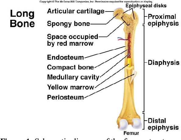
Figure (Huang et al., 2012)
Femur: Microscopic Description
Bone cells have a significant role in constructing bone tissues and the proper functioning of bones. Four types of cells are found in bone tissues which are “osteogenic cells, osteoclasts, osteocytes, and osteoblasts”. The diaphysis region consists of a compact bone (denser bone) lining. There is a hollow cavity in the diaphysis region that is filled with yellow marrow. On the other hand, the epiphysis region consists of spongy bone filled with red marrow. The meeting place of epiphysis and diaphysis is called “metaphysis”. (Florencio-Silva et al., 2015)
Femur: Functions
The femur connects the hip girdle to the knee joint, which gives the human body the liberty to move freely. This bone is known as the sturdiest bone in the body. The entire body structure is supported by the femur bone. It supports the entire body weight along with the movement of the body.
Femur: Homeostasis Balance and Imbalance
The homeostasis of the femur is maintained and balanced with the coordination of “osteoclasts, osteoblasts, and osteocytes” (Andersen et al., 2009). If these cells stop functioning properly, then the bone matrix will become unstable and will lose its homeostatic balance. The homeostatic imbalance in the femur can cause the bone to become weak and can cause a fracture.
Femur: Disease
“Legg-Calve-Perthes” is a bone disease that can occur in the femur. In this disease, the supply of blood to the proximal end of the femur gets interrupted, which is the source of bone weakness. If the blood supply gets disrupted for a long time, the bone starts to get weak, which can easily cause a fracture. (Legg-Calve-Perthes Disease – Symptoms and Causes, n.d.)
References
Andersen, T. L., Sondergaard, T. E., Skorzynska, K. E., Dagnaes-Hansen, F., Plesner, T. L., Hauge, E. M., Plesner, T., & Delaisse, J.-M. (2009). A Physical Mechanism for Coupling Bone Resorption and Formation in Adult Human Bone. The American Journal of Pathology, 174(1), 239–247. https://doi.org/10.2353/ajpath.2009.080627
Betts, J. G., Young, K. A., Wise, J. A., Johnson, E., Poe, B., Kruse, D. H., Korol, O., Johnson, J. E., Womble, M., & DeSaix, P. (2013). Bone Structure. https://opentextbc.ca/anatomyandphysiologyopenstax/chapter/bone-structure/
Bone cell biology: The regulation of development, structure, and function in the skeleton—Marks—1988—American Journal of Anatomy—Wiley Online Library. (n.d.). Retrieved December 8, 2021, from https://onlinelibrary.wiley.com/doi/abs/10.1002/aja.1001830102
Femur. (n.d.). Kenhub. Retrieved December 8, 2021, from https://www.kenhub.com/en/library/anatomy/femur
Florencio-Silva, R., Sasso, G. R. da S., Sasso-Cerri, E., Simões, M. J., & Cerri, P. S. (2015). Biology of Bone Tissue: Structure, Function, and Factors That Influence Bone Cells. BioMed Research International, 2015, e421746. https://doi.org/10.1155/2015/421746
Huang, B. W., Chang, C. H., Wang, F.-S., Lin, A., Tsai, Y., Huang, M. Y., & Tseng, J.-G. (2012). Dynamic Characteristics of a Hollow Femur. https://www.semanticscholar.org/paper/Dynamic-Characteristics-of-a-Hollow-Femur-Huang-Chang/cedb005d962397cddb10ee211f51fb6811629af6/figure/0
Human skeleton | Parts, Functions, Diagram, & Facts | Britannica. (n.d.). Retrieved December 8, 2021, from https://www.britannica.com/science/human-skeleton
Legg-Calve-Perthes disease—Symptoms and causes. (n.d.). Mayo Clinic. Retrieved December 8, 2021, from https://www.mayoclinic.org/diseases-conditions/legg-calve-perthes-disease/symptoms-causes/syc-20374343
Cite This Work
To export a reference to this article please select a referencing stye below:

Academic Master Education Team is a group of academic editors and subject specialists responsible for producing structured, research-backed essays across multiple disciplines. Each article is developed following Academic Master’s Editorial Policy and supported by credible academic references. The team ensures clarity, citation accuracy, and adherence to ethical academic writing standards
Content reviewed under Academic Master Editorial Policy.


